Local flow properties in the cardiovascular area can be linked to diseases and outcomes
Cardiac Flow Simulator (Particle Image Velocimetry)
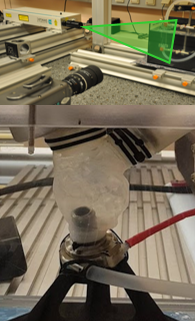
In a healthy heart, complex three-dimensional flow pattern vortices ensure efficient forward blood flow with reduced losses and swirling flow patterns. In transparent models of heart geometries these flow patterns can be studied and the effects of different pathologies can be analyzed. Changes in these vortex flow patterns can even be used as predictors for cardiovascular diseases.
The key technology used is Particle Image Velocimetry (PIV), which analyzes the effects of left ventricular assist devices, including variations in support, different positions, controlled speed modes and patient-specific heart geometries. In combination with the M3dRES infrastructure, possible links to diseases (e.g. thrombus formation, strokes) are investigated.
Computational Flow Dynamics (CFD)
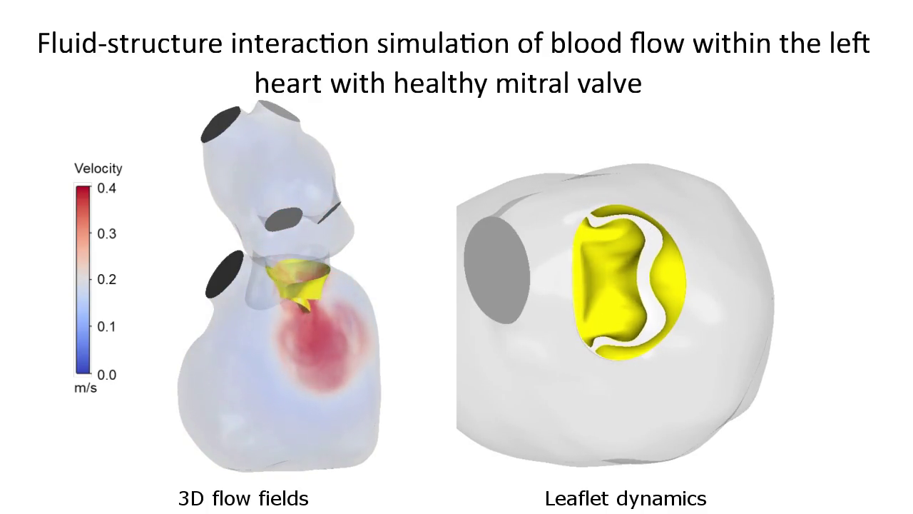
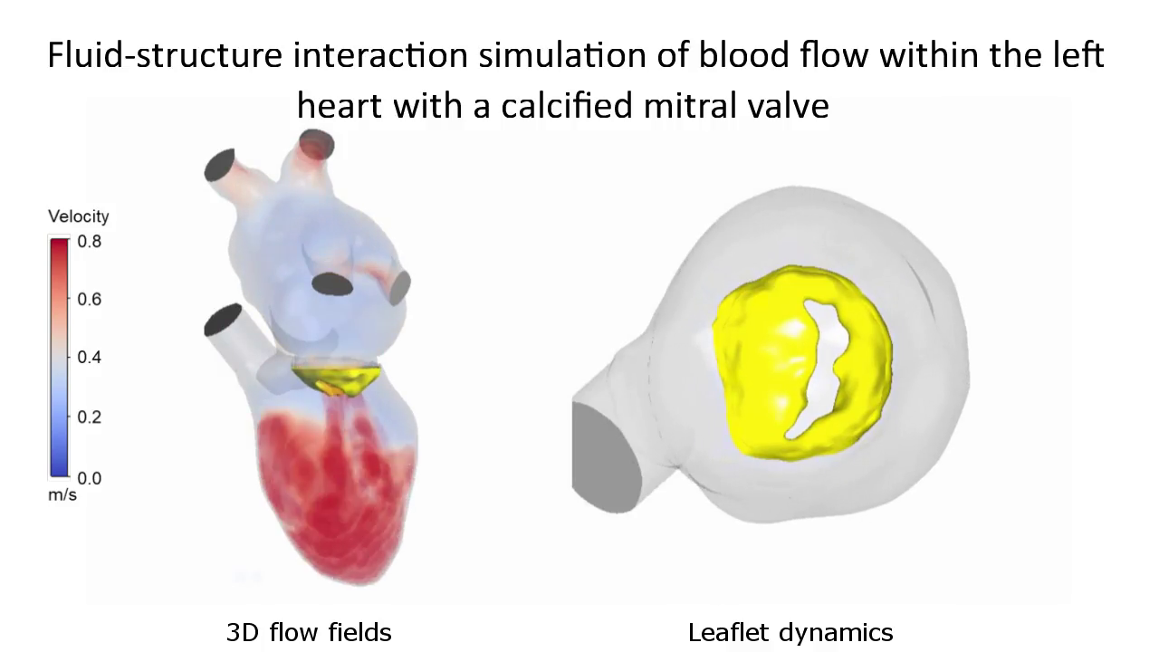
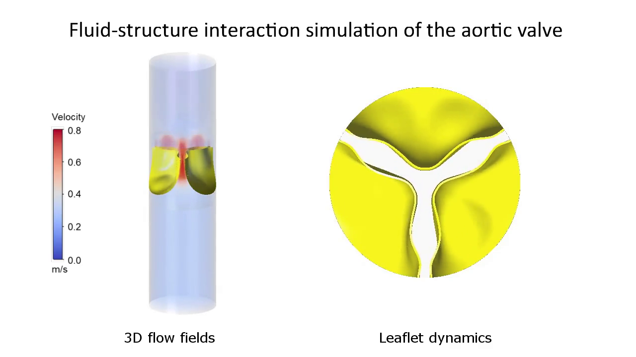
Parallel to the experimental flow studies, numerical methods are applied to gain further insights into cardiovascular flow. The general goal is to solve fluid mechanical problems approximately with numerical methods, which are performed at the Vienna Scientific Cluster (VSC). The numerical results will be validated against experimental measurements of different cardiovascular flows. Effects of different ventricle geometries, mechanical cardiovascular support, design of blood flow cannulas, heart valve are only some of the most burning research topics.
Patient-specific FSI simulation of intracardiac blood flow with a healthy mitral valve
After activation, data will be sent to YouTube. Further information here: Data protection
Patient-specific FSI simulation of intracardiac blood flow with a calcified mitral valve
After activation, data will be sent to YouTube. Further information here: Data protection
FSI simulation of the aortic valve
After activation, data will be sent to YouTube. Further information here: Data protection
Use of bioprosthetic material for simulators and training
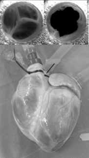
Bioprosthetic valves and tissues are a valuable source for patients with heart failure. These materials and additionally pig/bovine hearts or even just valves are a source of biological tissue that allows experimental investigations in mock loops and can help to reduce animal testing. Effects of different aortic valve replacements as well as valve pathologies can be created and used either for research purposes or as a training tool for cardiac surgeons or interventional radiologists.
Flowvisualition with Ultrasound-Techniques (Echo–PIV)
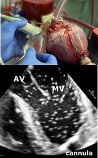
Cardiac echocardiography is the most important imaging routine in patients with heart failure and is also used to assess the performance of heart valves and heart muscle tissue. However, its reliability is sometimes unproven, especially with new devices. In the experimental setup, the performance of new devices or technologies (e.g. heart valves, anastomosis techniques) can be tested and evaluated using standardized experimental measurements (Particle Image Velocimetry - PIV).
