
Pioneering the development, design, and realization of biomedical imaging and sensing technologies with a focus on their clinical translation

Optical imaging involves a fusion of photonics, biology as well as (bio)medicine and deals with the interaction between light and biological matter. It offers tremendous opportunities for biotechnology development, fundamental as well as applied translational research. Merging of laser technology, photonics and biotechnology therefore sets new paradigms for non-invasive medical diagnostics.
Research Area Representative

Rainer Leitgeb
P +43 (0)1 40400-17140
Research Groups
Baumann & Merkle Group | Pircher Group | Leitgeb & Drexler Group | Rene Werkmeister | Zeiss Lab
More about this research area
Legacy
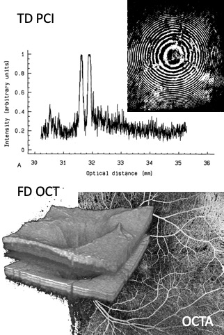
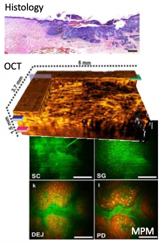
This Center is in particular pioneering in the field of Optical Coherence Tomography (OCT) as well as vascular laser diagnostics since the late 1980s (Prof. A. F. Fercher). Optical biometry as the first medical application of partially coherent laser interferometry has been successfully translated to clinical diagnostics. Furthermore, the principle of Fourier domain OCT has been pioneered at this center, which forms the heart of any modern OCT system today.
Optical Coherence Tomography Technologies
Based on more than three decades of experience, we are further pushing the limits of OCT technologies to meet the demands of tomorrow. New OCT technology developments comprise functional imaging, advanced contrasting schemes, resolution and speed enhancements, miniaturization (OCT on a chip), endoscopic probes, as well as advanced signal and image processing schemes.
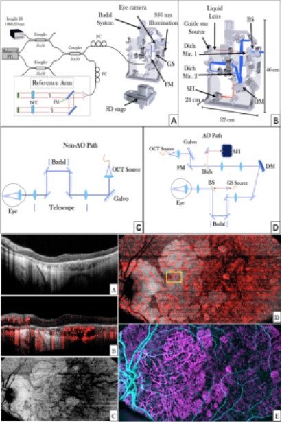
Translational Optical Coherence Tomography
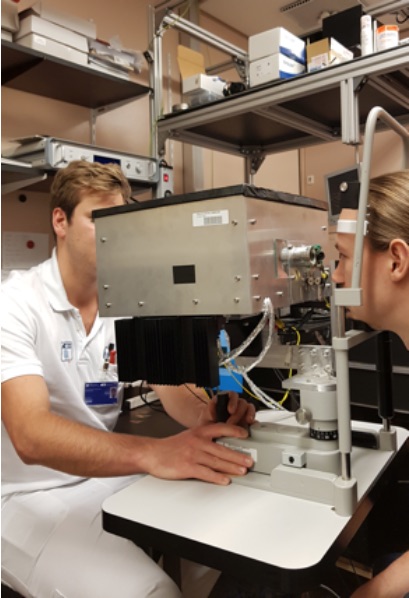
The translational activities embrace clinical and preclinical applications and are actively supported by partners from university clinics within the Medical University of Vienna, by external national and international experts, as well as by strong industry partners with an international network of stakeholders.
Photoacoustics
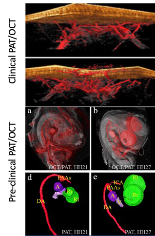
Optical imaging techniques are mostly confined to superficial lesions due to physical limitations of light propagation. Photoacoustics that is the optical excitation of an ultrasound wave in tissue, allows for deeper tissue penetration of up to a cm deep with close to optical resolution. This method uses absorbing endogenous chromophores in tissue, such as hemoglobin or melanin, but could potentially also target exogenous contrast agents.
Advanced Microscopy & Spectroscopy
Advanced light microscopy and spectroscopy provide a unique tool to explore structural, metabolic and molecular contrast in healthy and diseased tissue from small live animals (rodents, zebrafish, drosophila, and C. elegans) and biopsies. We have assembled a collection of wide-field fluorescence imaging microscopy, one-photon and two-photon laser scanning microscopy imaging, fluorescence lifetime imaging (FLIM), second harmonic generation (SHG) imaging, coherent Raman scattering (CRS) microscopy including coherent anti-Stokes Raman scattering (CARS) and stimulated Raman scattering (SRS) microscopy as well as spontaneous Raman scattering spectroscopy and imaging.
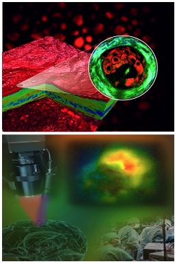
Multimodal Biomedical Imaging
By combining strengths of complementary imaging modalities it is possible to overcome limitations of the single modalities alone. OCT has a high structural sensitivity but lacks molecular sensitivity. The latter can be added through Raman spectroscopy, fluorescence and fluorescence lifetime imaging, photoacoustics, or through non-linear optical microscopy techniques. Functional imaging techniques such as OCT angiography enhance further the diagnostic strength of multimodal imaging. Multimodal imaging adds particular challenges for the design of imaging probes (endoscopes, laparoscopes, ophthalmic systems) across different scales.