Lumped-parameter modeling of cardiovascular hemodynamics and electrophysiology

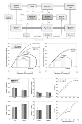
Mathematical models can help to gain a better understanding of human physiology and its changes associated with disease by providing a different perspective on its underlying mechanisms. Lumped-parameter models of the cardiovascular system including end-stage heart failure, heart failure with preserved ejection fraction, atrial fibrillation have been developed.
Recently, modeling has been utilized to predict cardiac responses to vagus nerve stimulation, thus helping better to understand the interaction between heart and pump, and to design pump controllers and future electroceutical therapies with the aim to restore vagal-cardiac control in heart transplant patients.
Development of novel neuromodulatory control strategies to restore vagal-cardiac control

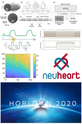
Cardiac vagal denervation following heart transplantation leads to reduced exercise tolerance and quality of life, potentially also to earlier graft failure. In this project a neuroprosthesis is developed to restore the vagal-cardiac closed-loop connection.
Closed-loop control helps to increase the selectivity of neuromodulatory devices while optimizing the response in the heart. However, due to the complexity and high non-linearity of physiological systems the design of such control strategies is very challenging. A model-based approach is pursued that helps to design and test novel neuromodulatory control paradigms while also minimizing the number of animal experiments. Part of this research has been funded by the EU-H2020 Program (NeuHeart Project).
Multimodal Nerve Imaging to desing intraneural nerve stimulation electrodes

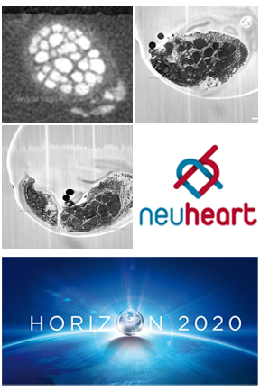
or the proper design of intraneural and epineural vagal nerve stimulation electrodes understanding of the nerve anatomy is required. A multimodal approach is used at our Center to image Cervical Vagus Nerve including cardiac branches.
Coherent Anti-Stokes Raman Spectroscopy and optical coherence tomography enable visualization and molecular characterization of myelinated nerve fibers and tissue structures (e.g. epineurium, fascicles). Micro-CT, delivers a detailed anatomical information of the Vagus, whereas the High Resolution Episcopic Miscrocopy produces 3D digital data histological tissue samples in high image quality. Part of this research has been funded by the EU-H2020 Program (NeuHeart Project).
