
Listening deep into tissue with molecular specific contrast
Photoacoustic Tomography
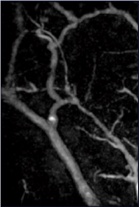
Photoacoustic tomography (PAT) uses diffusive optical illumination for the sample. The absorbers in the sample can then generate photoacoustic pulses. These pulses will be detected by the detector array. Using backpropagation image reconstruction, we can find the whereabouts of the absorbers inside the sample in 3D. We have performed photoacoustic tomography in both small animal imaging and clinical imaging. The image shows some human blood vessels imaged by the photoacoustic tomography system.
Photoacoustic Microscopy
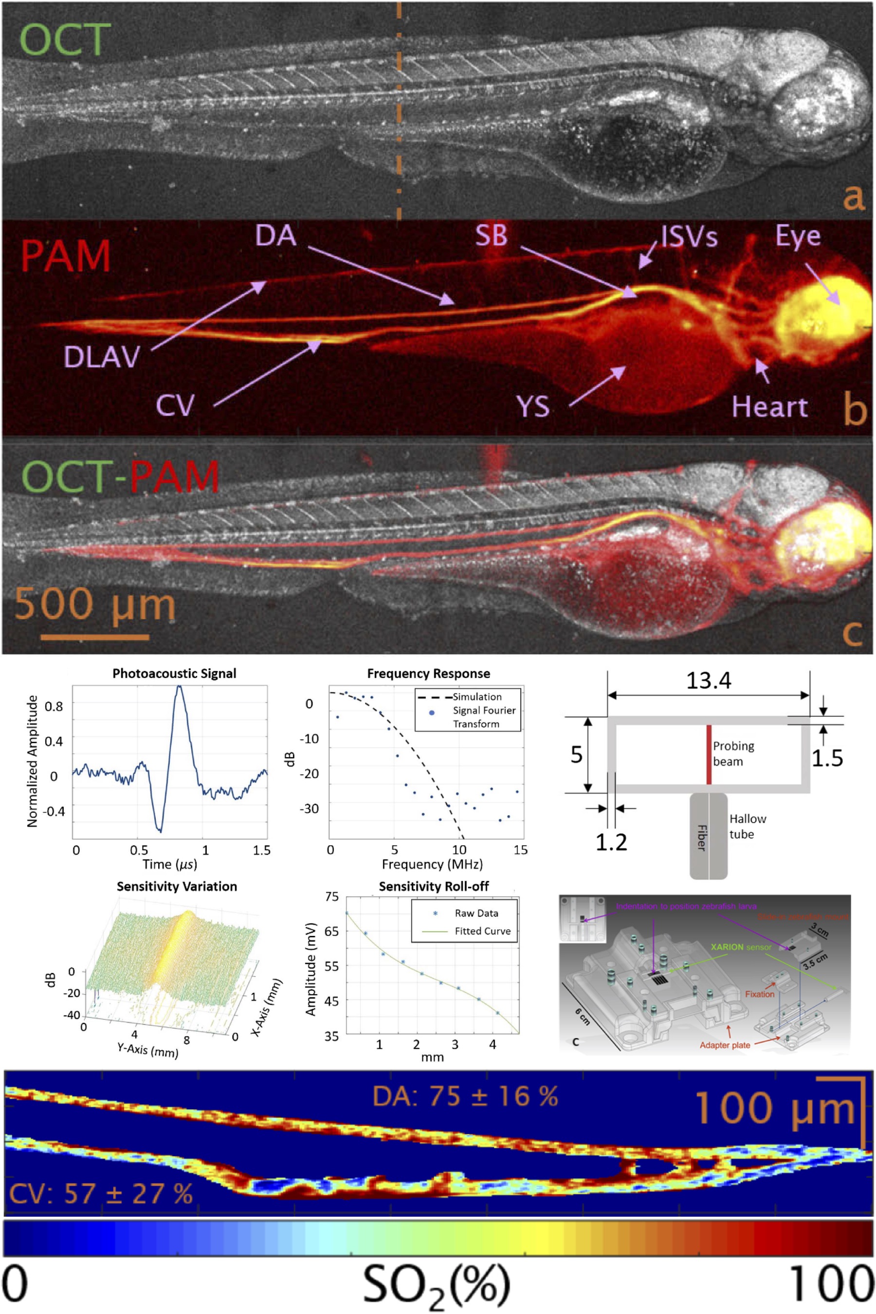
Optical resolution photoacoustic microscopy (OR-PAM) is ideally suited for pre-clinical small animal imaging. The sample (e.g. a zebrafish embryo) is illuminated with nanosecond pulsed laser light of various distinct narrow band wavelengths between 532 nm and 620 nm. The laser beam is optically focused and raster scanned along the sample. Therefore, absorbers can be located with a very high precision. Functional parameters, such as blood oxygenation, can be extracted in-vivo. A dual modality image of a zebrafish embryo is shown on top, while the zebrafish mounting concept and characterization of a novel akinetic acoustic sensor is shown in the middle. The bottom image describes the local oxygen saturation in a zebrafish embryo’s tail.
Translational Photoacoustics
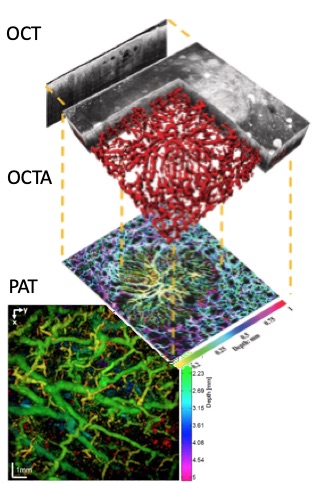
Translating the experimental findings to the clinics and preclinics is an important objective of our activities. In particular we combine the high structural sensitivity of Optical Coherence Tomography with the absorption contrast of PAT. Vascular details from superficial capillaries are seen with OCT Angiography, which are complemented by deeper and larger feeding vessels visualized with PAT. Clinical applications focus on dermatology. See more on multimodal imaging concepts.
Photoacoustic laparoscope (PAL)
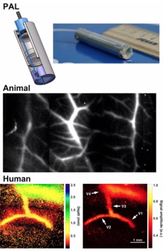
Photoacoustic imaging (PAI) can be a promising imaging tool for laparoscopy because the it provides label-free, high-contrast vascular imaging ability, which would benefit the diagnosis of diseases characterized by excessive angiogenesis, such as tumor and endometriosis. Its 3D imaging result is an inherent intraoperative guide for surgeons. Combining with molecular targeted contrast agents, PAI has the potential to further augment disease imaging to the cellular and molecular level.
Building on a close cooperation with industry and clinicians, a multi-spectral PAL is expected to deliver fast, label-free, 3D vascular imaging for laparoscopy.
Photoacoustic Elastography
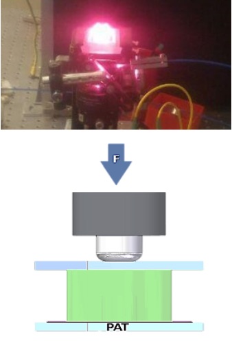
Diseases like arteriosclerosis or cancer are often accompanied by micro-scale stiffness changes in the affected area. Photoacoustic is able to provide 3D microscale images, which deliver together with a small compression force enough information to determine these biomechanical parameters, which can be used to find and characterize physiological and pathological tissue in and ex vivo. In combination with optical coherence tomography even more precise results can be achieved. The image shows photoacoustic imaging in action and a scheme describing photoacoustic elastography.