
A unique tool to explore structural, metabolic and molecular contrast in healthy and diseased tissue from small live animals and biopsies
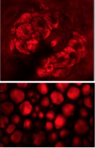
Fluorescence microscopy

Fluorescence imaging is the most rapidly evolving microscopy technique nowadays in the medical and biological field. Especially, fluorescence guided surgery has had an immense impact on the neurosurgical routine within the last two decades.
Second harmonic generation imaging microscopy

Second harmonic generation (SHG) is a parametric nonlinear processes providing structural and molecular information that is complementary to two-photon excited fluorescence (TPEF) imaging. A particularly strong attribute for biomedical applications is that SHG is able to visualize the tissue structure based on a purely endogenous type of contrast mechanism (label-free).
Coherent Raman scattering microscopy
Coherent Raman scattering (CRS) imaging techniques represent a powerful optical tool for biomedical diagnosis with coherent anti-Stokes Raman scattering (CARS) and stimulated Raman scattering (SRS) the most established among them. With these label-free imaging techniques based on vibrational spectroscopy, the spatial-temporal distribution of various molecules in complex samples, particularly biomedical specimens, can be investigated with the ability to visualize molecular composition and function in living systems.
Spontaneous Raman scattering spectroscopy & microscopy

Spontaneous Raman spectroscopy is widely applied to medical questions. Raman spectra yield a vast information about the molecular composition of tissue. Therefore, Raman spectroscopy can discriminate tissue types based on their molecular characteristics. Depending on molecular differences, healthy tissue exhibits a different Raman spectrum compared to pathological tissue such as tumors, making this technique extremely attractive for diagnostic purposes.