
Combining strengths of complementary imaging modalities to overcome limitations of single modalities alone
Raman/OCT Endoscopy
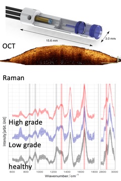
OCT successfully demonstrated its capability to reveal superficial tissue changes caused by early forms of cancer. This structural information can further be complemented by metabolic information on tumor grade gained by Raman spectroscopy. The synergistic combination of Raman and OCT imaging promises a fully non-invasive pathologic assessment of early stage tumors, which is critically needed for immediate treatment decisions. Within a European consortium, we are developing multimodal forward viewing endoscopes for bladder cancer diagnosis to be translated to the clinics.
Raman/OCT Retinal Imaging
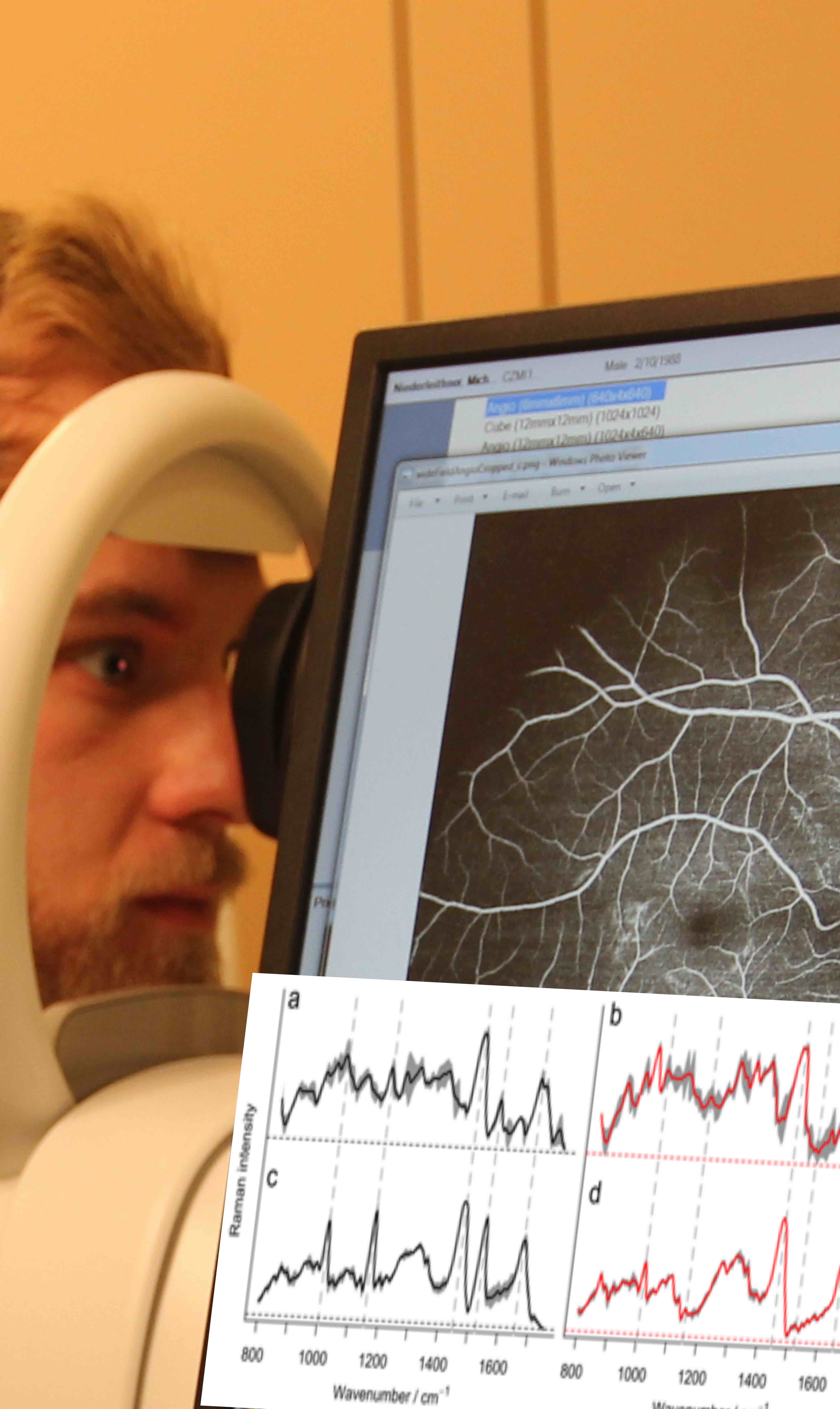
Molecular imaging of the back of the eye is expected to reveal early changes of retinal metabolism which could be precursors of diseases. For many diseases, early diagnosis allows to start treatment when it is most successful and before critical irreversible neural tissue loss has occurred. Furthermore, the human retina is often regarded as window to the brain and as such may give access to neurological disorders such as Alzheimer’s in a fully non-invasive manner.
OCT/OCTA/PAT
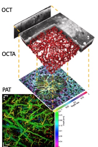
The combination of OCT and non-invasive, label free OCT angiography has been successfully applied to dermatology. However, the penetration of OCT is limited to about 2 mm into the skin. Photoacoustic tomography (PAT) on the other hand penetrates several millimeters deep into skin and reveals larger vascular structures. The combination of OCT and PAT therefore yields a more complete volumetric picture of perfusion and its alteration down to several millimeters into skin.
OCT/NLOM
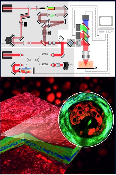
Non-linear optical microscopy (NLOM) techniques deliver images on the cellular level with metabolic contrast. The molecule specific functional information of NLOM complements the structural information obtained with OCT. A challenge is the light delivery with short pulses for NLOM through optical fibers as is for example needed in endoscopes. Hollow core fibers promise to overcome limitations of conventional optical fibers for efficient light excitation and collection.
OCT/PAT Endoscopy

The deep penetration of PAT promises to enhance the diagnostic capability for assessing tumor/ suspicious lesions not only in dermatology, but also for inspecting inner organs. For non-invasive assessment of the esophagus wall, this is achieved by multimodal tethered endoscopes that can be swallowed.