Development of advanced OCT technologies for biomedical imaging
Functional imaging

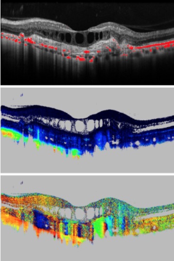
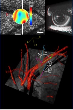
Functional imaging technologies are developed to analyze tissue function, in addition to obtaining the structural information of conventional OCT. These technologies comprise, among others, OCT angiography and Doppler OCT for qualitative and quantitative imaging of blood flow, spectroscopic OCT to measure parameters like tissue oxygenation, and optophysiology, a method that measures and quantifies tissue response to an optical stimulus by OCT signal analysis.
Contrast enhanced imaging

Conventional OCT records images based on the backscattered signal intensity. These images frequently do not provide a direct differentiation of tissues.
Contrast enhanced OCT imaging exploits other properties of light to provide additional image contrast: Polarization sensitive OCT can differentiate fibrous tissues by their birefringence and depolarizing tissues by their scrambling of the light’s polarization state. Multi-directional OCT exploits the anisotropic scattering properties of some tissues for contrast generation, and spectroscopic OCT uses the spectral dependence of the attenuation coefficient for tissue differentiation and for quantitative measurements.
High-resolution imaging

The resolution of commercial OCT systems is limited by the bandwidth of the light source (in axial direction) and by the numerical aperture (NA) of the imaging system (in lateral direction).
For improved axial resolution, light sources with very large bandwidth can be used, e.g. Ti:Sapphire lasers and supercontinuum sources. With these sources, axial resolutions down to 1 µm and even lower can be achieved. For imaging of the retina, the lateral resolution is limited by the aberrations caused by the ocular media which only allows imaging with low NA. Adaptive optics OCT systems that correct for the ocular aberrations enable retinal imaging with cellular resolution.
OCT on a chip

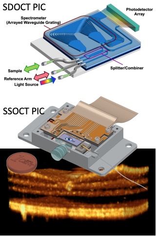
Photonics integrated circuits (PIC) promise a paradigm shift in OCT with respect to size, costs, and portability. OCT core systems comprising light source, interferometer, and sensors fit on a chip of the size of a cent coin. For spectrometer based OCT (SDOCT), compact PIC based spectrometers have been implemented. Still, the line array would consume larger space. Swept source based OCT (SSOCT) would be the most compact solution, as balanced sensors can be easily integrated on the chip together with compact light sources. The challenge is to overcome optical coupling losses to and from the chip. The developments are pushed by strong European project consortia from academia and industry led by Medical University of Vienna.
Endoscopic imaging

Endoscopes allow OCT to penetrate also deeper into the human body to reach inner organs. Especially for cancer diagnosis, OCT provides important diagnostic information about tumor stage and to distinguish non-cancerous lesions from early stage tumor. The combination with label-free 3D OCT angiography (OCTA) further enhances the diagnostic potential. MUW collaborates with IMTEK, University of Freiburg, who contribute expertise for the endoscope design and implementation. Applications range from urology, gastroenterology, to surgical guidance in neurology. A complete image on tumor pathology is targeted by multimodal endoscopic OCT.
OCT signal and image processing

Several gigabytes of data are acquired in very short time during a single 3D image acquisition with modern high-speed OCT systems. In clinical trials, hundreds of patients are imaged, leading to huge amounts of data. The rapid online display of images, as well as the post-processing and evaluation of acquired data require rapid processing algorithms and artificial intelligence methods. Modern technologies like GPU based data processing and neural networks are developed to support scientists and clinicians to analyze the results and to identify and evaluate novel biomarkers for precision medicine.
Research Groups
Baumann & Merkle Group | Pircher Group | Leitgeb & Drexler Group | Werkmeister Group | Zeiss Lab
