Development of an anthropomorphic Phantom to evaluate and adjust image quality in Digital Breast Tomosynthesis
What is Digital Breast Tomosythesis?

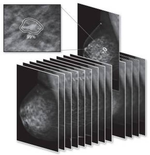
Digital breast tomosynthesis (DBT) is a pseudo 3D modality for early detection of signs of breast cancer. It is designed to improve the detection of suspicious structures in comparison to (2D) full-field digital mammography (FFDM) by reducing lesion masking of the normal breast anatomy and overlaying structures. The clinical value of DBT in diagnostics and breast cancer screening in comparison to FFDM was already investigated and various observer performance studies have demonstrated that DBT as stand alone modality or in combination with other diagnostic techniques increases cancer detection in comparison to single FFDM. These results led to an increase of the number of DBT systems in clinical practice.
Prototype of an anthropomorphic test phantom

The main characteristics of the phantom are: its 3D outer shape that is half of cylinder (200 mm diameter, 48 mm height), the 3D background structure created with PMMA spheres of different diameters submersed in water and lesion like models in different sizes that can be tested for detectability. The difference in attenuation coefficient between PMMA and water is close to the difference between adipose and glandular tissue.
The current phantom has 4x five clusters with microcalcifications with increasing diameter from 90 µm to 250 µm, non spiculated lesions based on real lesion models from a database (from 1.6 mm to 6.2 mm) and spiculated lesions (always the same model printed with different sizes from 3.8 mm to 9.7 mm with spicules on the surface).
Diagnosis of Mammography Images with Artificial Intelligence (AI) Software
In a pilot project together with Braincon (iCAD representative for Austria) and Radiologicum Margareten (Screening- and Assessment Unit in Austrian Mammography Screening Program), we evaluate diagnostic detection rates of ‘Profound AI’ in comparison with results from radiologists. Consequently, the CAD Software from Hologic will also be evaluated and a direct comparison of both AIs is planed.
Clinical Performance Benefits of ‘Profound’ from previous studies
Based on FDA reader study results, the software offers radiologists:
- 8,0% increase in sensitivity
- 6,9% average increase inspecificity
- 7,2% average reduction in recalls
- 5,7% average improvement in radiologists AUC
- 52,7% reduction in reading time

Workflow Benefits
- AI algorithm with superior performance for the detection of breast cancer and high specificity with low numbers of false positives
- Certainty of Findings and Case Scores to assist in clinical decision-making and prioritizing caseloads
- Workflow solution that renders quick results while analyzing each image for detections
- Deep Learning technology allows for continuously improved algorithm performance via updates
