
Increasing spatial resolution on an MR-scanner for human and biomedical applications
Hard-/software development and quality control for MR-microscopy
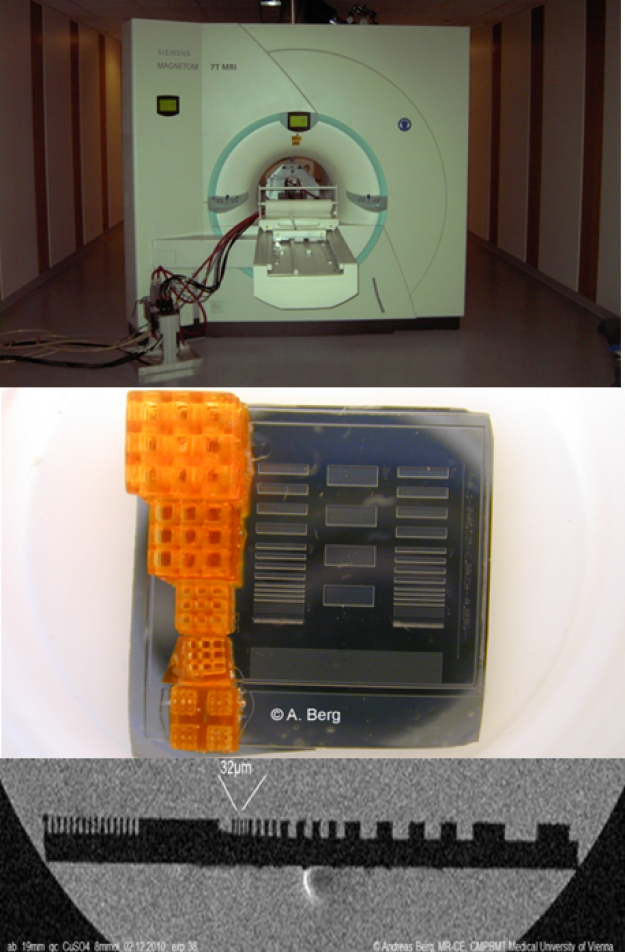
Using dedicated prototype hard- and software the spatial resolution on High-field MR-scanners for human MRI-diagnostics can be significantly increased up to the microscopic level (pixel size < 100µm). The high spatial resolutions allow in principle for the early and more detailed diagnosis of pathologies ex-and in-vivo on humans but also research on biological model systems.
We implemented a strong microscopy insert and several sensitive radiofrequency-sensors on the patient layer of a 7T MR-scanner designed for human applications. For quality assessment specifically designed test objects (phantoms) have been developed to check quantitatively the spatial resolution via modulation transfer functions in 2 and 3 dimensions.[Berg, Deutsches Patentamt, DE19904635]; [Berg et al. Proc. ISMRM 2010, progr nr. 1048]; [Berg et. al., Front. Phys. 2023].
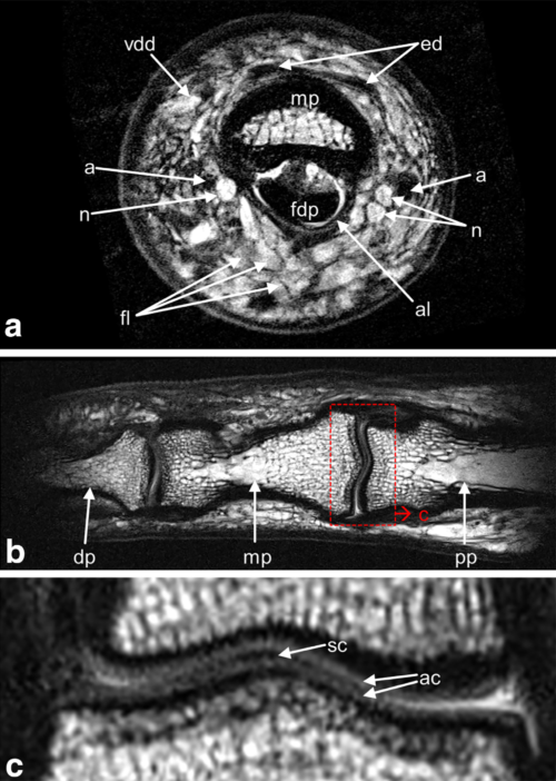
Broadline/UTE-imaging of rigid tissue and biocompatible materials with fast signal decay (e.g. tendons)
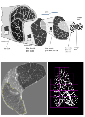
There are several tissues in the human body (e.g. tendon, meniscus, teeth) which cannot be visualized with inner structure due to the very quick signal decay. Ultrashort encoding time (UTE) methods along with MR-microscopy are capable of detecting uniquely morphological substructure, e.g. the self-similar tissue anatomy of the tendon (fig. left). Even the fractal dimension of the self-similar tissue texture can be evaluated using differently contrast weighted MR-microscopical data and data-processing software. [Berg et al. Proc. ISMRM 26, 2831 (2018)].
Alternatively, polymer based objects can be visualized by MRI using add-ons to the polymer-Matrix during the manufacturing process [Rausch et. al. European Patent Office publication server 2022].
Applications of MR-microscopy
MR-based histology
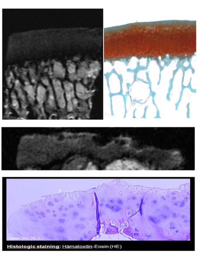
Though MR-microscopy is not capable of achieving the high resolution of optical microscopy multi-slice multi-contrast images at about 30x30µm2 pixel size can be obtained, which is sufficient for the detection of e. g. osteoarthritic changes in the cartilage. The main advantage of differently parameter weighted MR-microscopy is represented by the non-invasive imaging process without the necessity of cutting the sample into thin slices. Also some staining processing might be avoided as there are several different contrast weighing mechanisms in MRI available (e.g. sd, T1, T2, T2*, susceptibility, DWI, MT,..) [Berg et. al. Proc. ECR EPOS #2914 (2011)], [Hager et. al., JMRI, 2022].
MR-based polymer gel dosimetry
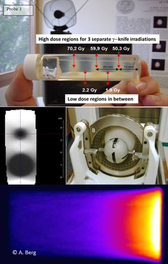
In radiation therapy for tumor treatment a critical problem is related to the dosimetry of small sized photon or particle beams (proton therapy) due to the relative big size of ionization chambers and the extensive measurement time for a complete volume data set. Multi-slice (3D) dosimetric images can be obtained by T2-Parameter selective imaging (T2-maps). T2-µ-imaging allows for high resolution 3D-dosimetry of the smallest radiation fields for tumor therapy in clinical routine e.g. with γ-knife (middle image). Also the visualization of mm-sized dose Bragg-peaks at the final end in particle therapy (bottom) is possible [Khan et. al. Polymers 11(10): 1717 (2019)].
Radio frequency (RF) coils for MR-microimaging on humans
Highly sensitive in-house built RF coils allow for micro-imaging, e.g. of human extremities in-vivo on an ultrahigh magnetic field MR-scanner (7T). Using a dedicated finger coil it is possible to depict the internal structures of the human finger in-vivo within patient‐compatible measurement time. Various anatomical features such as bone and marrow, tendons and annular ligaments, cartilage, arteries and veins, nerves and Pacinian corpuscles can be visualized at pixel size of about 80×80 µm². This may serve for diagnosis and treatment monitoring in pathologies ranging from inflammatory or erosive joint diseases to injuries of tendons and ligaments to nervous or vascular disorders in the finger (Laistler et al. Magn Reson Med 79:588–592, 2018).