
We develop and use various MR imaging and spectroscopy techniques to investigate human tissues and diseases. Here we summarise our work activity in the thorax and abdomen. Physiologic motion, such as heartbeat, circulation, breathing or peristaltic activity, has to be taken into account during MR scans which often last for several minutes.
Based on our history, our strength is in spectroscopy, in particular phosphorus, carbon and proton spectroscopy, in investigating energy metabolism. Due to breathing motion in the thorax and abdomen we have developed sophisticated navigator techniques to tackle these challenges and to increase the sensitivity of spectroscopy measurements. We currently extend our efforts to ambitious imaging projects.
Cardiac MR spectroscopy

After activation, data will be sent to Vimeo. Further information here: Data protection
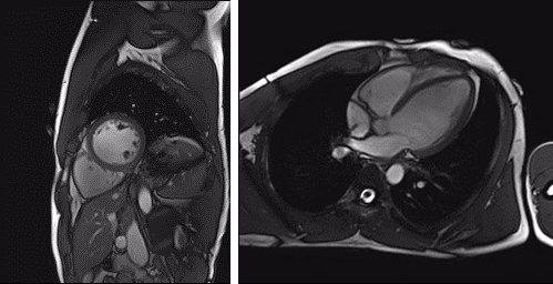
The heart is responsible for keeping us going and thereby consumes considerable energy itself. As a pump, and being situated in between the lungs, it is constantly in motion, which poses challenges to imaging and to capturing its metabolism by MRS. Cardiac energetics and respiratory motion are therefore our current central topics of investigation.
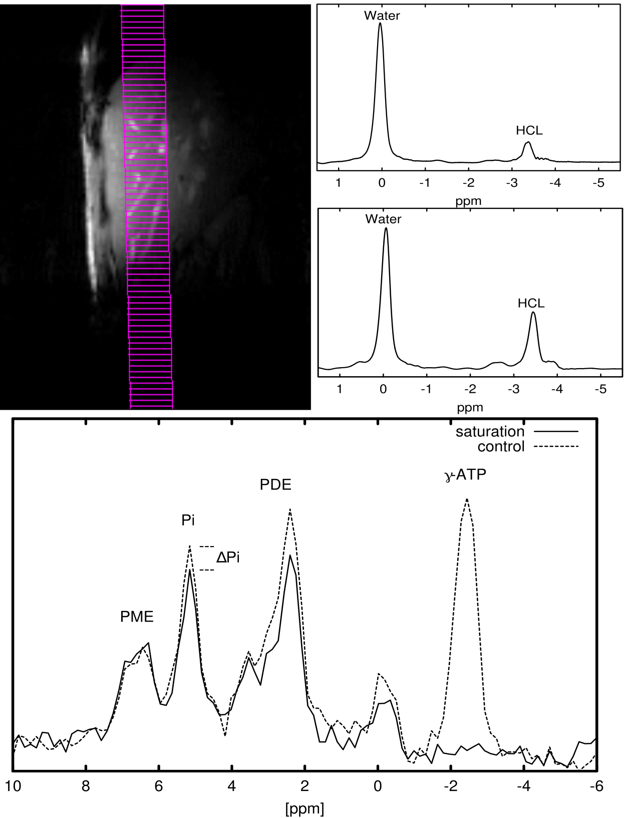
Here we apply our expertise of studying skeletal muscle metabolism. The thin layer of myocardium enclosing the ventricles imposes challenges in SNR and in separating the signals. We use multi-nuclear interleaving for image-navigated ³¹P MRS to improve localisation accuracy.
Motion compensation using MRI navigators

Breathing motion is a big issue in cardiac magnetic resonance imaging and spectroscopy. External trackers, such as respiratory belts or even optical cameras can only deliver indirect indication concerning the actual motion of the organs in abdomen. Specialized rapid MRI scans, so-called MR navigators, offer a unique opportunity to gain sufficient information on the motion of internal organs.
We have developed 3D heart detection and tracking approaches for both 3T and 7T scanners, which allow to void uncomfortable and unreliable prolonged breath hold for detailed cardiac MR acquisitions.
Renal imaging
Chronic kidney disease is a severe condition affecting one in ten people, which leads at end stage to a lifesaving need for dialysis and transplantation. Associated healthcare costs of this global burden are estimated to be more than 1 trillion USD worldwide.
The main challenge is that at early stages affected individuals are often unidentified in the clinics due to the limitations of blood and urine samples. Together with European and international partners (www.renalmri.org) we are optimising non-invasive MRI techniques to improve diagnosis of chronic kidney disease.
Liver metabolism
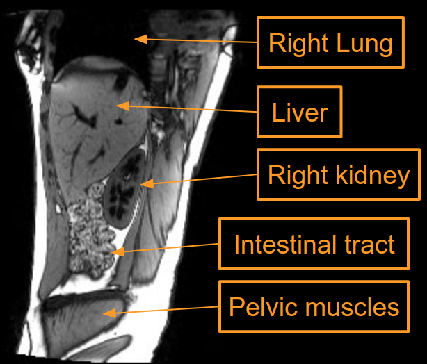
The liver is the central chemical plant of our body. Nutrients and toxins from the intestines are filtered, metabolised or stored for later release. It is no surprise that the role of the liver is central in the pathophysiology of many diorders.
Disease-induced metabolic alterations often precede structural changes by years and are a direct window to tissue function. In particular, hepatic lipid, glycogen and high energy phosphate contents and their turnover are very sensitive markers.
Motion detection using external sensors

For rigid objects and non-deformable organs the deteriorative effects of motion on MRI data may be entirely undone if the accurate position and orientation information is available in real time. Optical motion tracking systems are capable of delivering such information with a sufficient accuracy and a low temporal latency.
Several systems have been developed to track the position of specialized markers, while being exposed to strong magnetic and radio frequency fields. One or several cameras located in the magnet bore have proven effective for accurately tracking head motion during long acquisitions and thereby improving the image quality in long MRI acquisitions.
Research Groups
Meyerspeer Group | Schmid Group | Laistler Group | Frass-Kriegl Group