
Dynamic multi-nuclear MR Spectroscopy and Imaging in vivo open unparalleled opportunities to study energy metabolism and perfusion in skeletal muscle.
Skeletal muscle phosphate kinetics
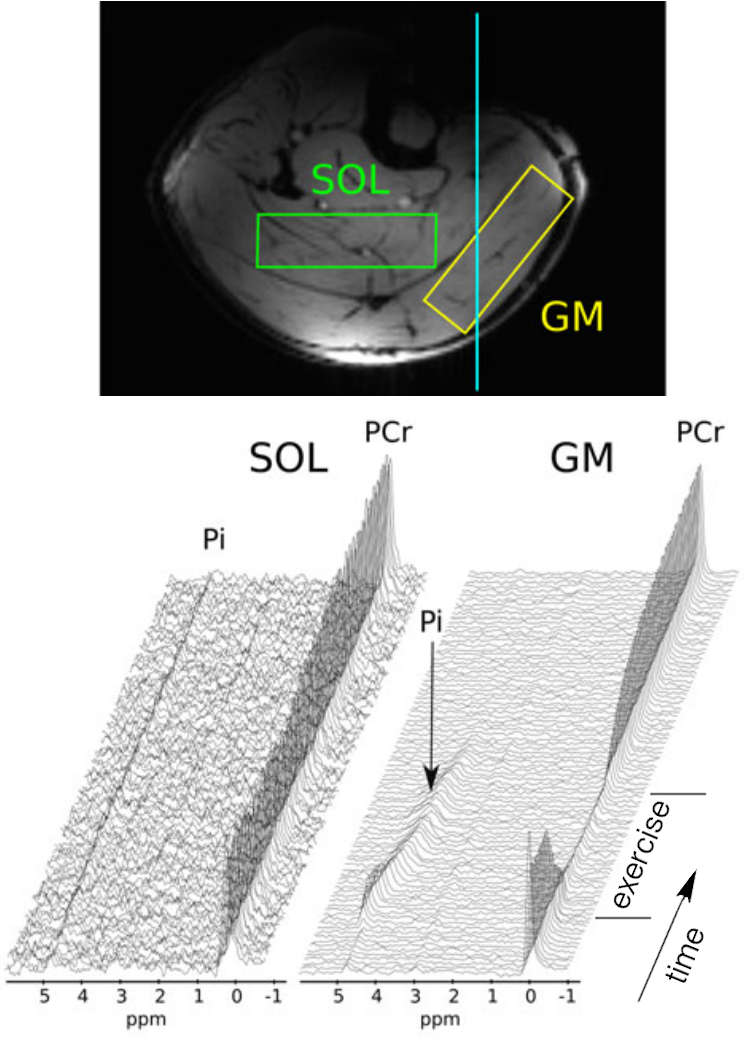
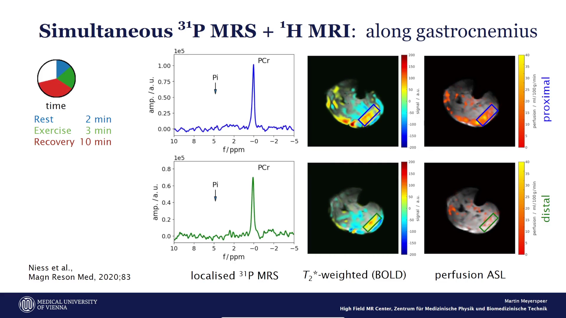
Skeletal muscle can ramp up its energy turnover by orders of magnitude. Exercise recovery during ³¹P MRS is uniquely suited to study cellular mitochondrial function, glycolysis and proton kinetics in situ. We develop and combine imaging and spectroscopy techniques and dedicated RF coils to locally measure ATP, phosphocreatine (PCr) and pH directly in the exercising muscle in real time.
After activation, data will be sent to Vimeo. Further information here: Data protection
Skeletal muscle perfusion
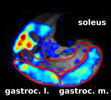
Blood flow and capillary perfusion deliver nutrients and oxygen to the muscle and help dispose metabolic products. Perfusion deficits or ischaemia can cause severe disorders and lead to immobilsation, infection and reduced healing. Transient ischaemia can be induced safely by pressure cuffs. Perfusion rates can change dramatically between these impaired states and maximal exercise in healthy tissue. Arterial spin labeling (ASL) tags and traces arterial blood spins. Our developed sequences generate perfusion data every few seconds allowing time-resolved measurements without external contrast agents, simultaneously with metabolic information from spectroscopy.
1H and 13C MRS of skeletal muscle
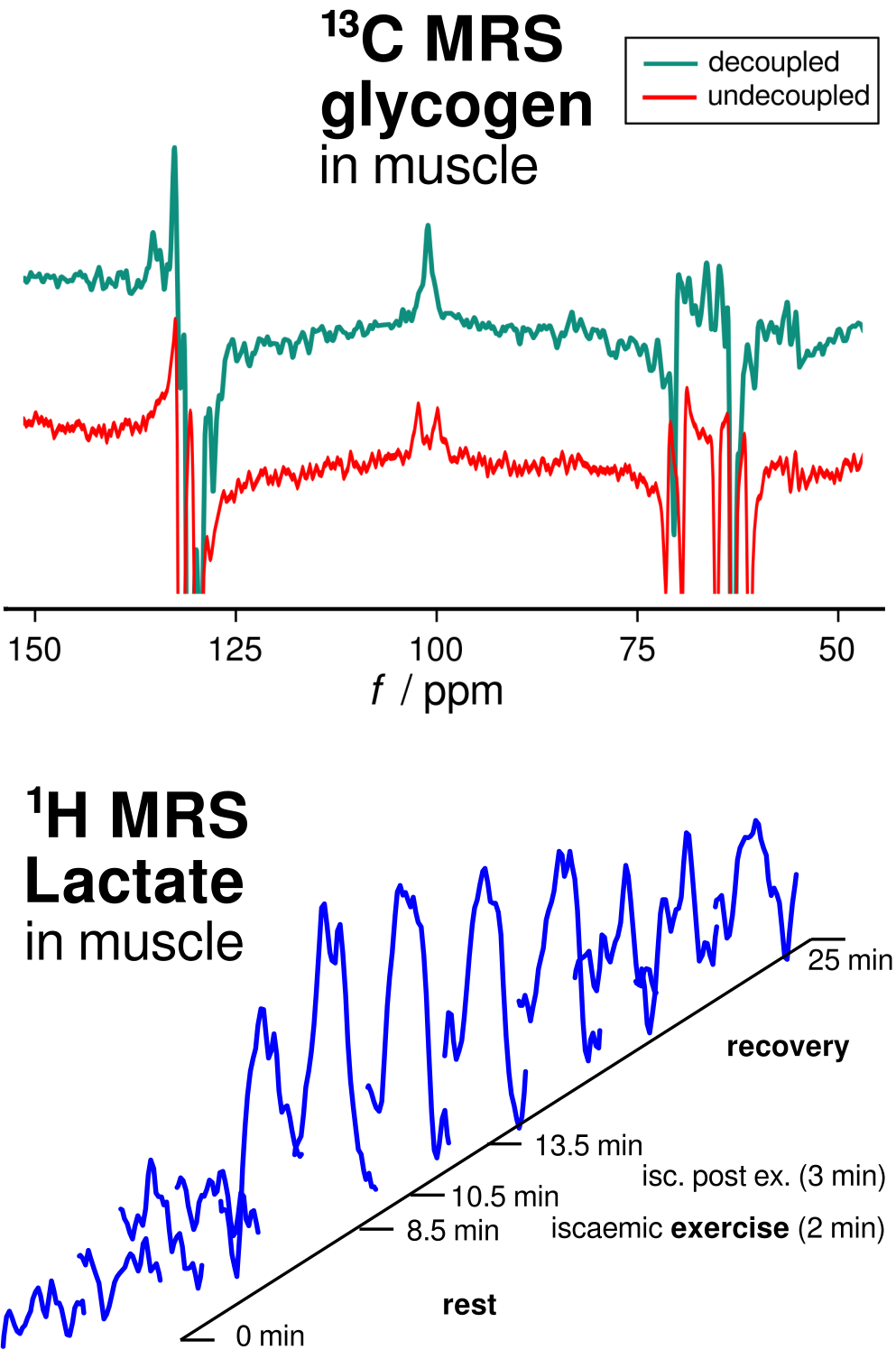
Glycogen is stored in muscle tissue as an energy reservoir. Its concentration can be quantified non-invasively by 13C MRS at natural abundance, i.e. without administering labelled substances. Proton decoupling can be applied to increase the sensitivity of measuring the small 13C MR signals, by collapsing the multiplet resonances into singlets.
Under high work load and in anaerobic conditions, glycogen, glucose and other metabolites can be converted to lactate, which temporally accumulates in the muscle cells. Measuring lactate non-invasively, directly in the muscle, requires special spectral editing techniques like double-quantum filtered 1H MRS. These measurements can complement the metabolic information from 31P MRS.
We have investigated and applied measurements of intracellular muscle fat (IMCL) which are sensitive markers of insulin sensitivity, impairment of which is a precursor and feature of diabetes mellitus.