
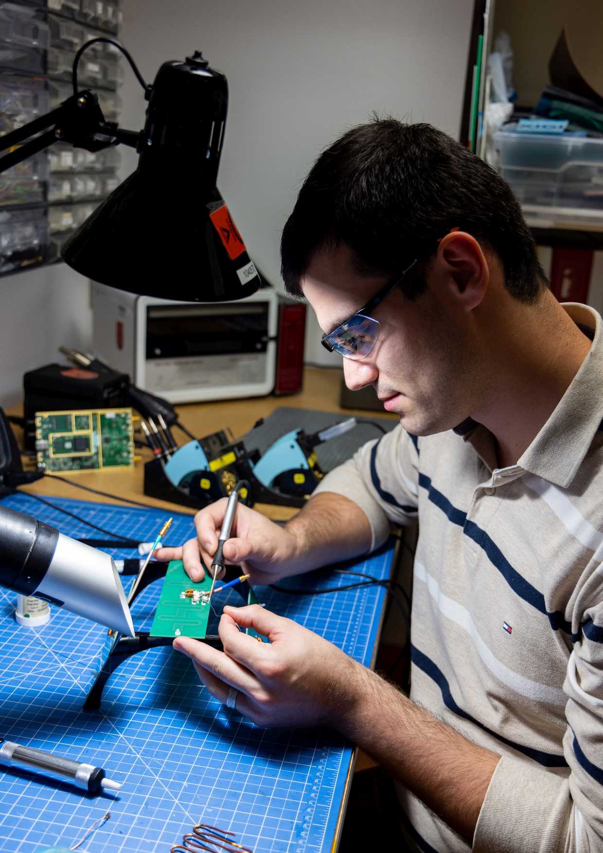
RF coils are the key hardware for acquisition of the MR data and strongly determine the achievable data quality. They are used for excitation of the nuclear spins in the body as well as for receiving the resulting MR signal.
In our RF Lab, we develop RF coils with the goal to enable new MR applications and explore novel design approaches towards wearability and flexibility. Increased specificity, in particular for metabolic studies in the muscle or the body, is achieved by multi-nuclear coils. Coils for multi-modal applications enable the combination of MRI and other technologies like PET and TMS. Small and particularly sensitive coils can be used for MR microimaging and microscopy. Dedicated coils for animals accommodate the specific requirements for animal studies. While designing new coils, we develop and investigate novel concepts of coil construction and functionality.
Flexible Coils
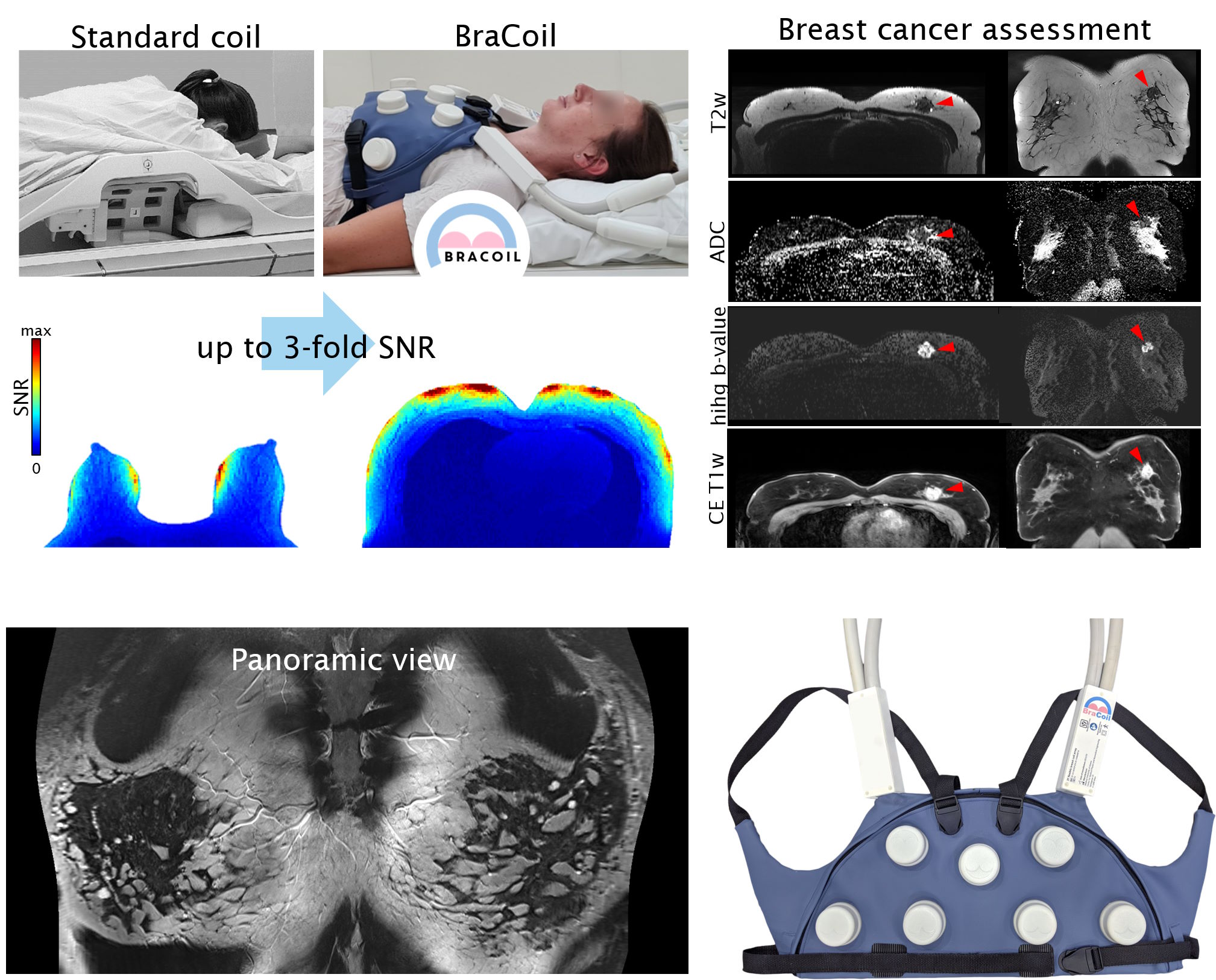
3T BraCoil: a wearable coil vest for breast MRI
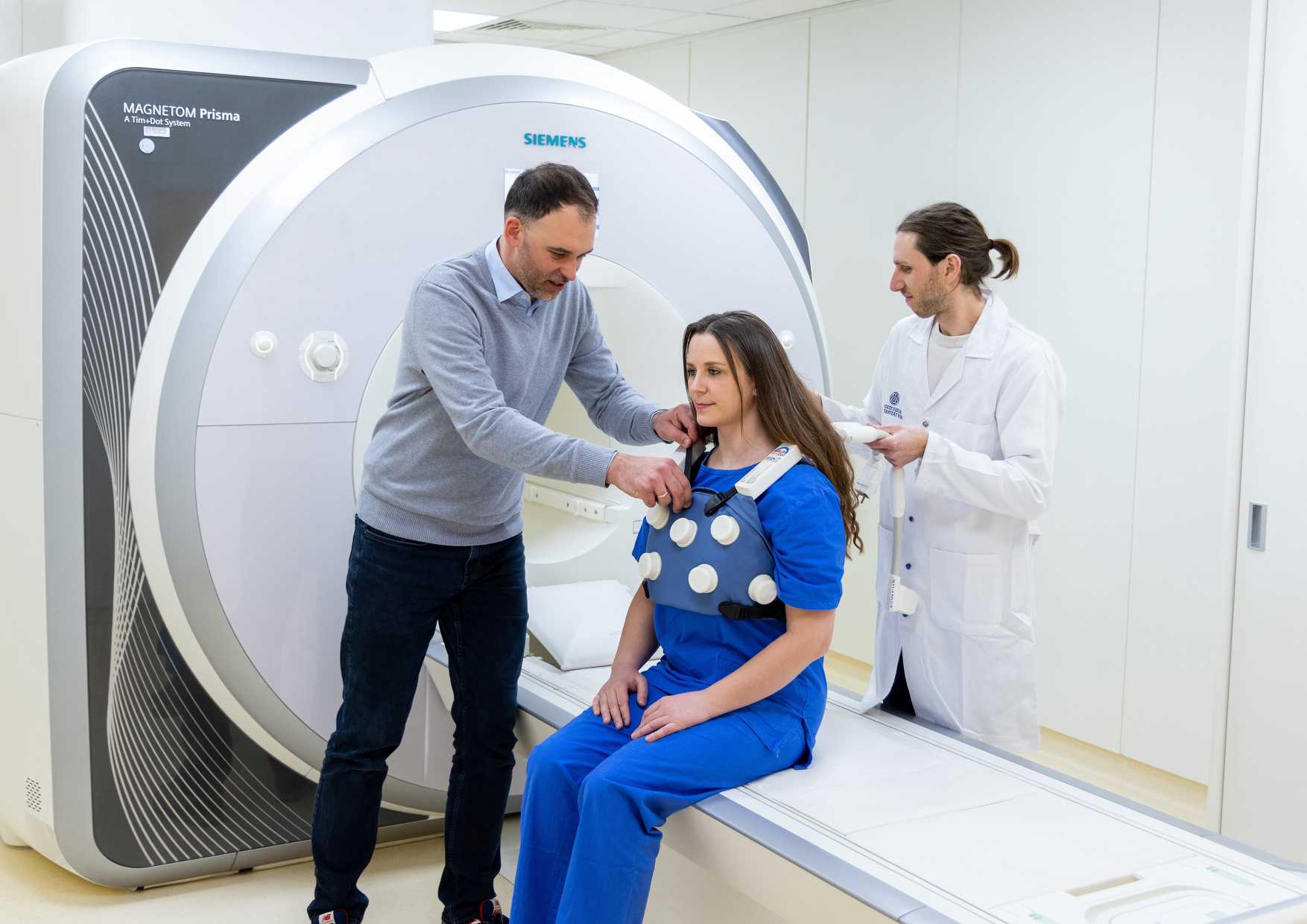
Based on ultra-flexible coil elements we developed a 28-channel coil array that can be worn like a vest, which increases patient comfort in breast cancer examinations and results in excellent image quality with up to 3-fold signal-to-noise compared to state-of-the-art breast coils. Current breast coils require the patient to lie face down on the patient bed and have their exposed breasts hanging into openings of the coil, which are too large for small breasts and too small for very large breasts. The BraCoil is always tightly fit to the surface of the breast regardless of size, and therefore delivers high signal for any breast size. The BraCoil can be used in supine (=face up) position, in which the breast shape is similar to ultrasound exams or surgery. This allows for easier localization of lesions and facilitates the clinical workflow, saving time, money, and potentially unnecessary additional exams.
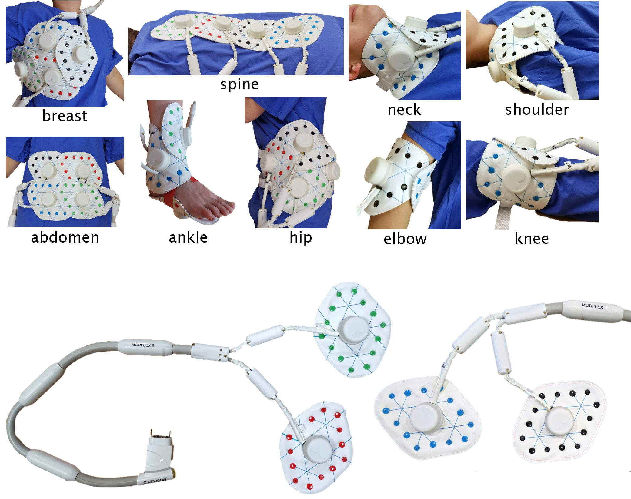
3T ModFlex: modular flexible coil array system
We designed a novel coil array system called "ModFlex" for magnetic resonance imaging (MRI) using flexible, lightweight 4-channel coaxial coil modules. These coils are used to acquire high-quality images of various body parts. In a study we compared ModFlex with traditional MRI coils in six different anatomical regions: neck, ankle, spine, and hip. Results showed that ModFlex outperformed or had similar performance compared to traditional coils. This means faster and higher-resolution MRI scans are possible, especially in ankle and spine imaging. ModFlex is versatile and accommodates different patient sizes, potentially enhancing patient comfort during MRIs, making it a promising advancement in medical imaging technology.
3T flexible 23-ch array coil
We developed a flexible, application-ready 23-channel RF coil array. The array is composed of conventional loop coils fabricated on a rigid-flex printed circuit board. Lumped components needed for coil interfacing are placed on the rigid parts of the PCB covered by robust housing. Flexible PCB parts in between assure the mechanical flexibility of the device. This coil is suitable for high-resolution MRI of various anatomical targets, e.g. hand, ankle, knee, shoulder, with its most prominent application being MRI of the occipital cortex.
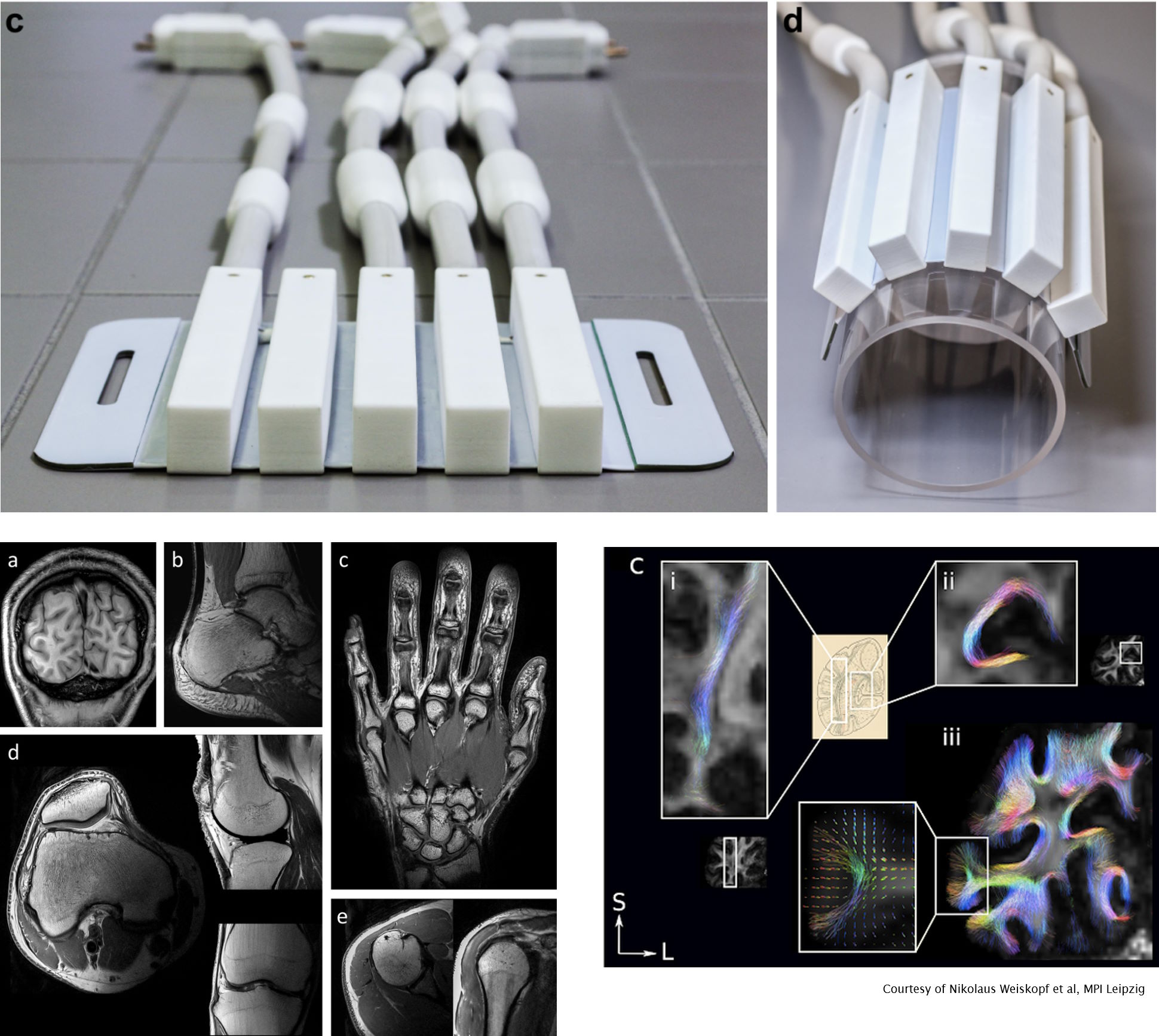
7T flexible 31P/1H cardiac coil array
To obtain both anatomical (1H imaging) and metabolic (31P spectroscopy) information from the heart, we developed a flexible array conforming to the chest shape based on transmission line elements for 1H and flexible wires for 31P. A considerable amount of electronic circuitry needed to be fit into the 3D-printed housings: power splitter, transmit/receive switches, tuning and matching networks, cable traps, preamplifiers. We used fullwave 3D electromagnetic simulations to determine the optimal phase setting between the transmit channels.
Multi-nuclear coils
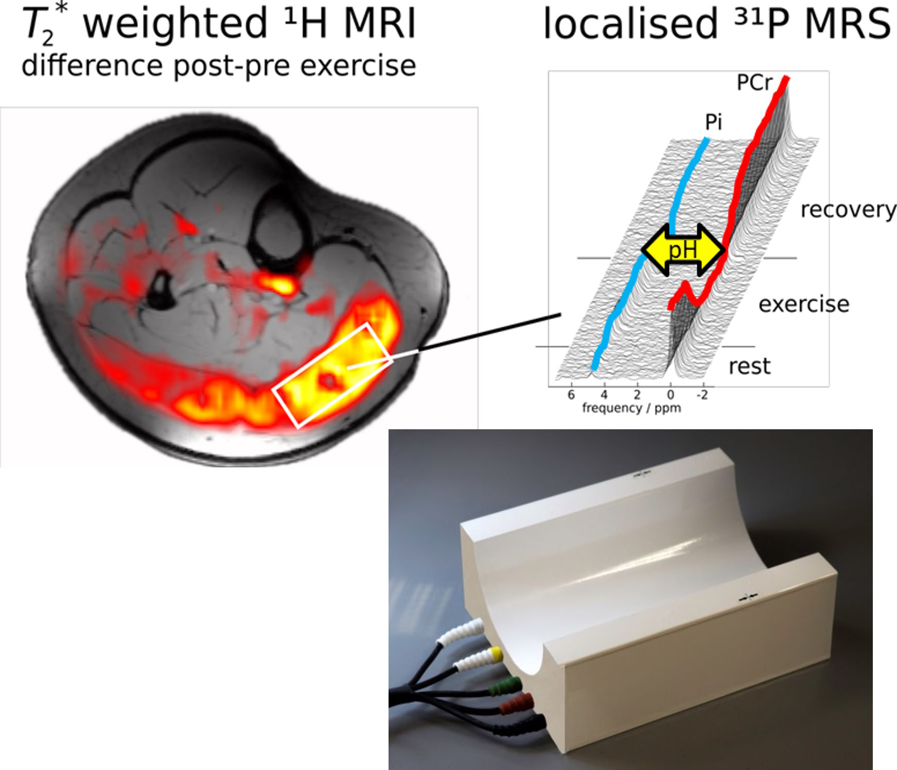
7T 31P/1H calf muscle coil
This half cylinder-shaped coil has 2 channels for 1H imaging and 3 channels for 31P spectroscopy (and imaging) and is designed to enable the detection of the metabolism of high-energy phosphates in the calf muscle. Numerous studies in muscle physiology have been performed with this coil, given its superior signal-to-noise ratio.
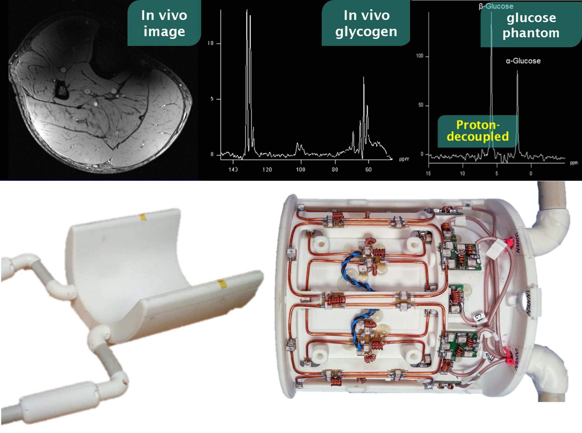
7T 13C/1H calf muscle coil
Carbon-13 spectroscopy can provide insights into sugar metabolism in the body via the measurement of glycogen and glucose. Due to the nature of the 13C MR signal, these signals appear split into different frequencies, hampering data analysis. This can be overcome by transmitting at the 1H Larmor frequency while detecting the 13C signal, a technique known as “proton decoupling”. To the coil builder, this means that extremely efficient filters (~100 dB) need to be included in the coil to avoid crosstalk between the strong 1H transmit signal and the subtle 13C MR signal. The coil we developed fulfills this criterion and provides four transmit/receive 1H channels combined with three transmit/receive 13C channels.
Multi-modal coils
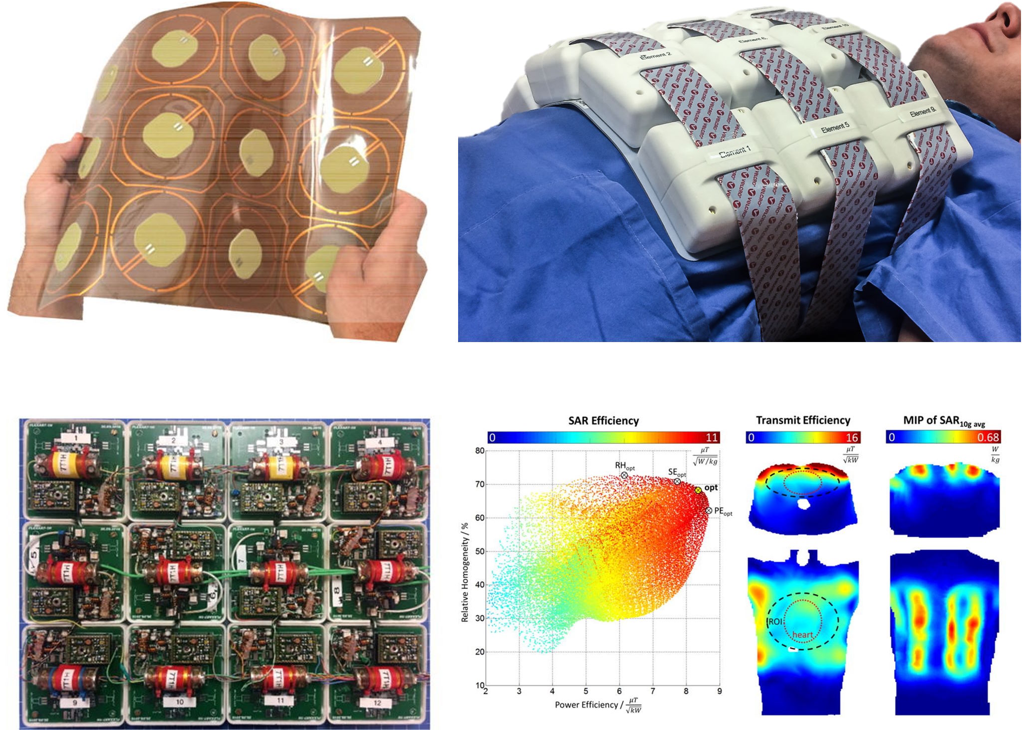
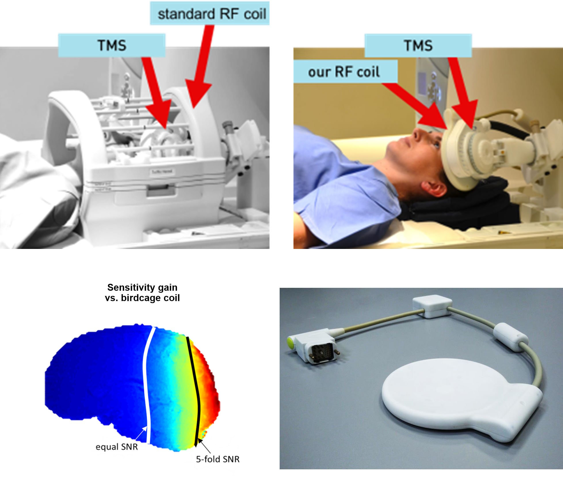
3T fMRI/TMS array coil
Transcranial Magnetic Stimulation (TMS) is a technique that uses strong pulsed magnetic fields to evoke brain stimulation in the area underneath the stimulator device. Using functional MRI (fMRI), this stimulation can be visualized. Traditionally, the TMS device had to be put inside a large birdcage-type head coil, which has the disadvantages of low MR image quality, slow acquisition, and very restricted possibilities for placement of the TMS device. Our approach was to develop a very thin RF coil array that fits between the TMS device and the head. This enables drastically improved MR image quality and very flexible positioning of the TMS device, at only a minimal loss in TMS stimulation efficiency due to the slightly increased distance of the TMS to the head.
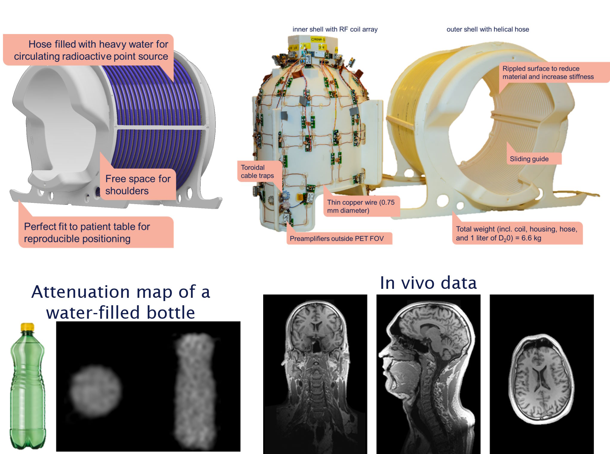
3T MR/PET head+neck coil with integrated attenuation correction
Positron Emission Tomography (PET) relies on radioactive tracers accumulating in cancerous tissues, emitting photons at 511 keV when decaying. On their way out of the body, they are attenuated by the surrounding tissues. To correctly quantify the PET signal, the spatial distribution of the attenuation coefficient has to be accounted for, which requires a map of the attenuation coefficient. Our coil includes a solenoidal hose through which a pellet with radioactive tracer can be pumped at a controlled speed, allowing for a transmission measurement of attenuation. The RF coil is designed in a way to minimize attenuation within the field of view of the PET system and covers the whole head including the neck.
Microscopy coils
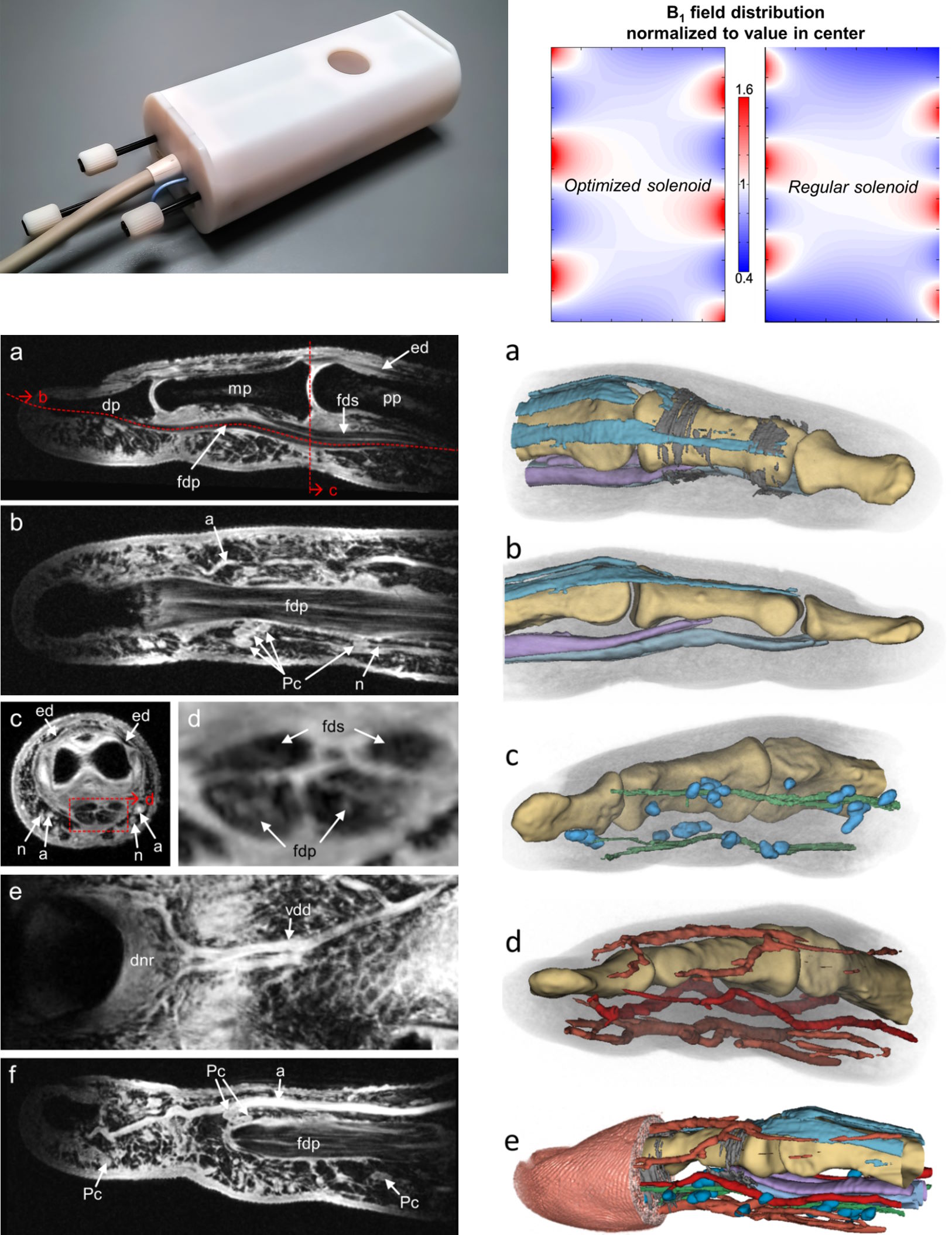
7T finger coil
To visualize the small structures of the human finger, we developed a solenoidal coil with optimized winding steepness to improve signal homogeneity. Using this coil, images with 100-200 µm³ isotropic resolution made it possible to segment and visualize bones, cartilage, tendons (including their internal structure), pulleys, arteries and veins, nerves and Vater-Pacinian corpuscules, as well as fat and skin.
7T microscopy array
Our 7T scanner is equipped with a microgradient insert providing a gradient strength of 750 mT/m and an inner bore diameter of 90 mm, which allows for high resolution imaging. We developed an 8-channel 1H receive array that tightly fits into an existing 72-mm diameter transmit birdcage coil, leaving a remaining bore size of 34 mm.
1.5T and 3T skin imaging and superconducting coils
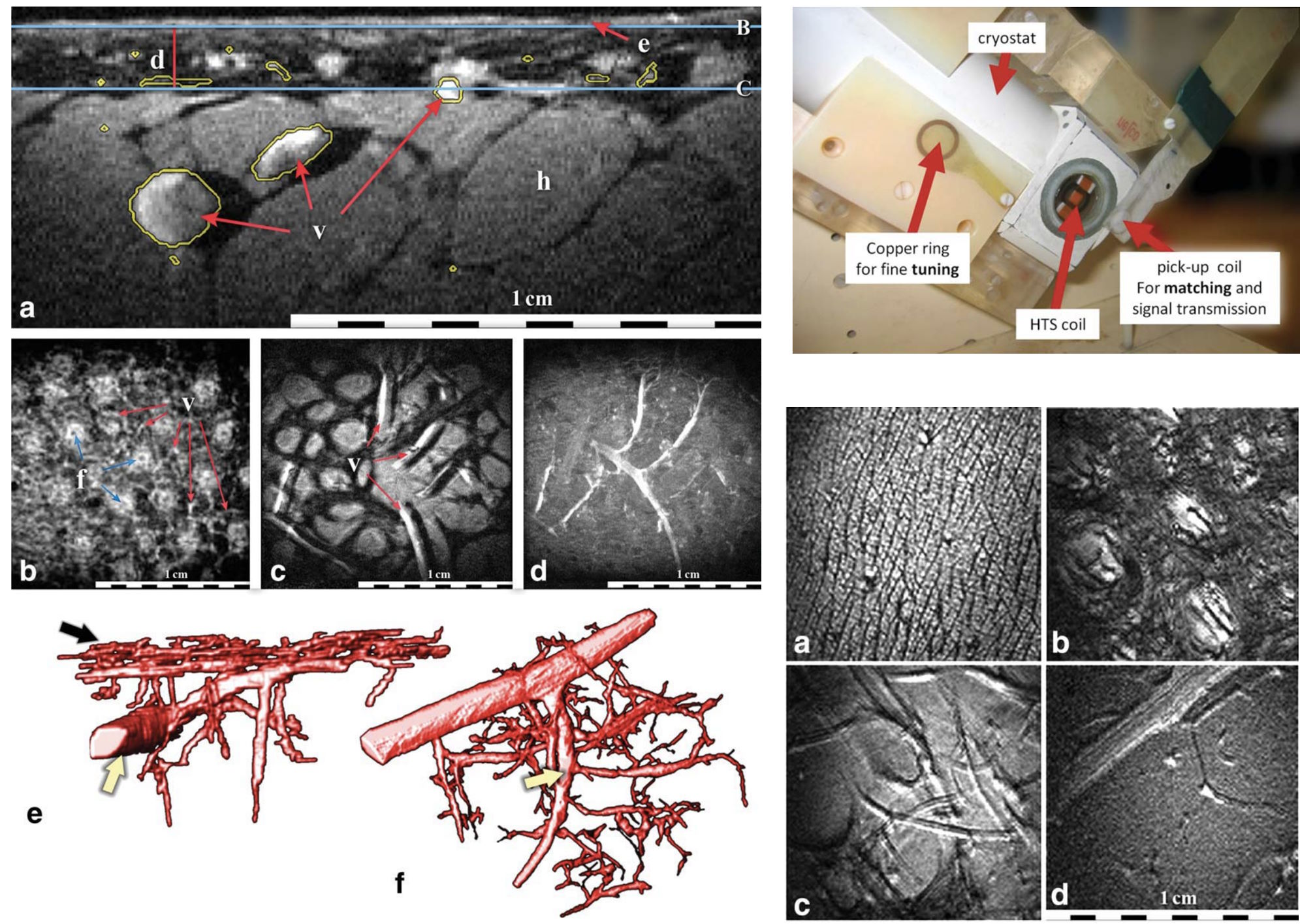
Very small surface coils can be used to achieve high signal-to-noise ratio close to the body surface, which allows for high resolution imaging of the human skin, enabling for example the depiction of small vascular structures in the dermis and hypodermis. When reducing the coil size, however, normal conducting coils produce more noise than the noise coming from the tissue, which limits image quality. With our collaboration partners in Paris we therefore used superconducting coils to obtain images of down to 20 µm³ isotropic resolution.
Coils for animals
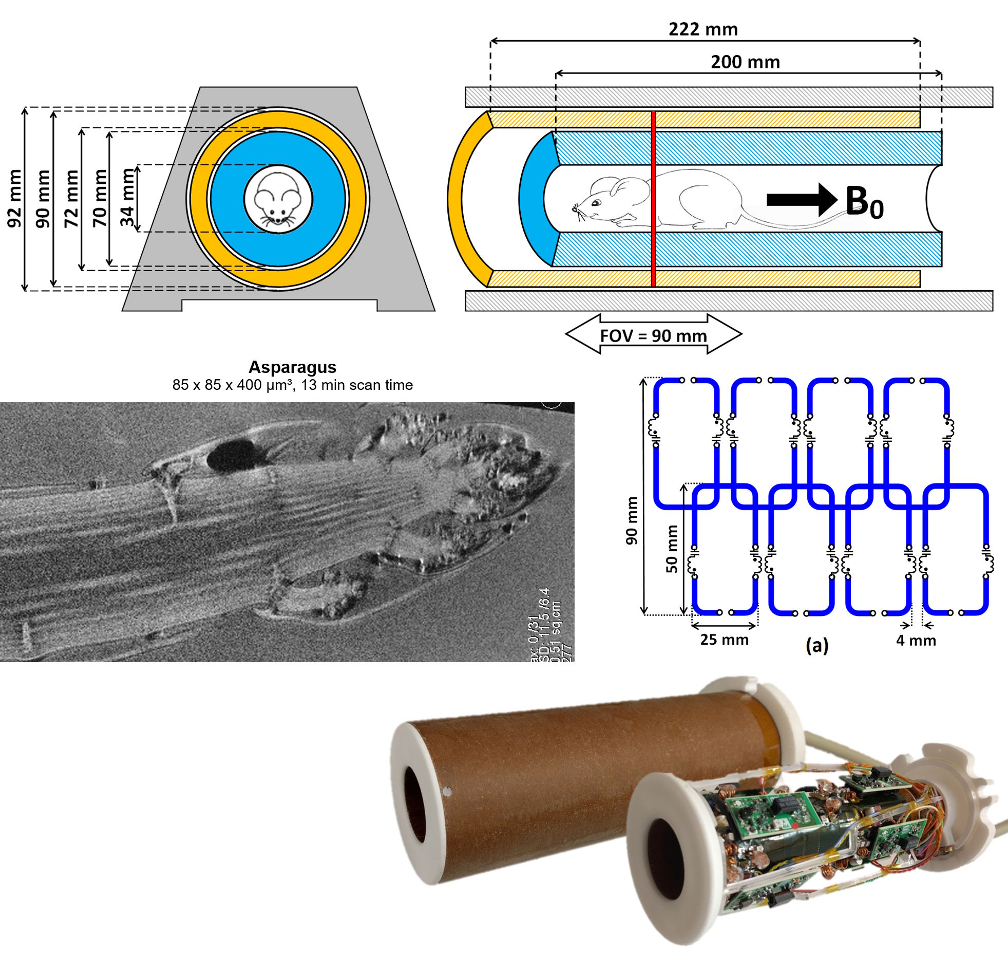
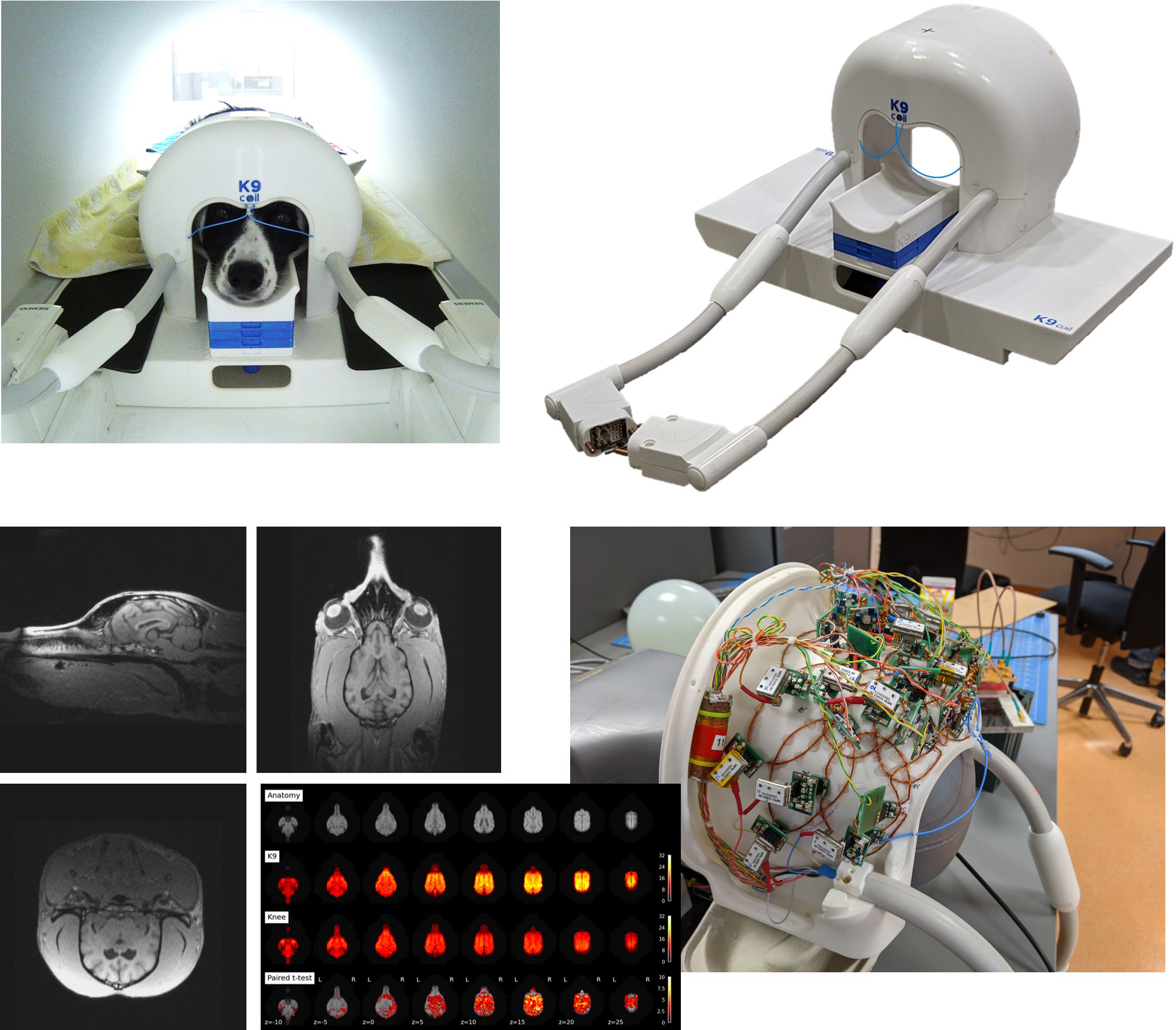
3T canine (K9) coil array for dog fMRI
Our collaborators at the Messerli Institute/Veterinary University of Vienna have trained dogs to voluntarily go into an MR scanner and lie still for a functional MRI examination in a behavioral study. We have developed a dedicated coil for this purpose. To obtain high quality images and make the exam as comfortable for the dog as possible it has a chin rest adjustable in position and height and the housing shape is designed so that the dogs can place their paws either under or next to the coil. The coil array is conformed to an average Border Collie’s head, with coil element sizes being smaller close to the brain (on top of the coil) and getting larger towards the sides with increasing distance.
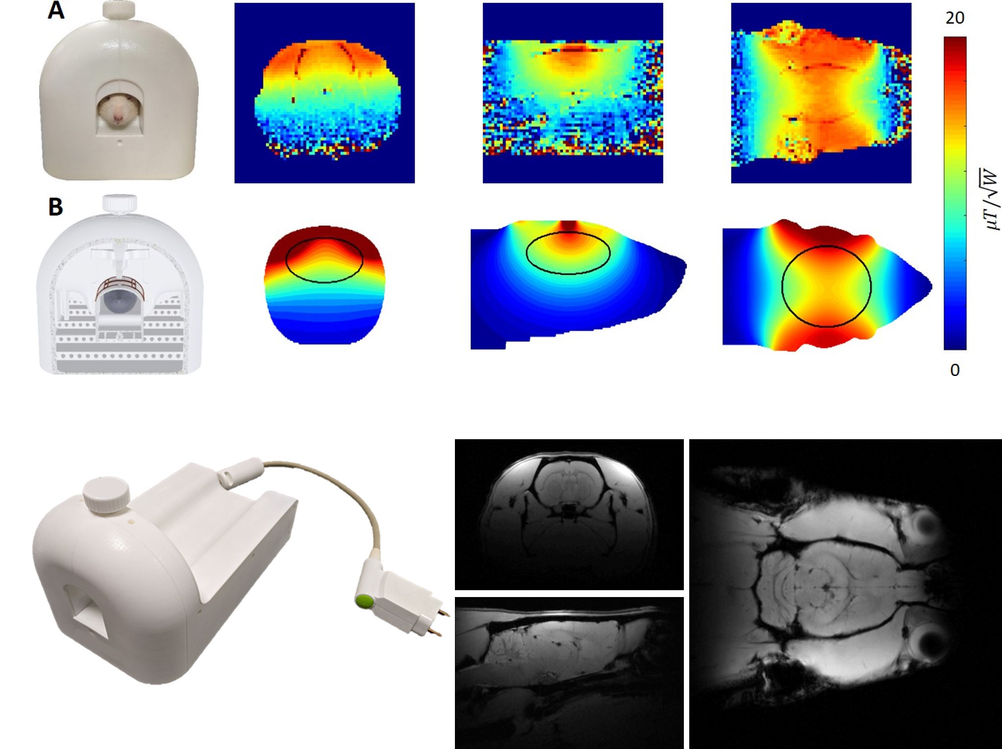
7T rat brain coil
We developed a 4-channel transceiver coil for rat brain imaging in a human whole body 7T scanner. The coil layout was optimized using electromagnetic simulation to achieve highest efficiency. As a special feature the height of the coil array can be adjusted to perfectly fit rat heads of various sizes from juvenile to large adult rats.
Novel coil concepts
Flexible multi-turn multi-gap (MTMG) coaxial coils
Our group is invested in fundamental research exploring the potential of novel coil technologies for improved imaging sensitivity in MRI. We introduced Multi-turn Multi-gap Coaxial Coils (MTMG-CCs), offering flexibility and form-fitting capabilities. MTMG-CCs allow adjustment of coil diameter when operating the coil at self-resonance equal to the Larmor frequency at 3 T and 7 T, making it possible to optimize imaging for specific body parts. The study also includes an equivalent circuit model to predict the resonance frequency of these coils. Through experiments and simulations, we found that MTMG-CCs perform comparably to standard coils while offering better patient comfort, paving the way for wearable MRI coils.
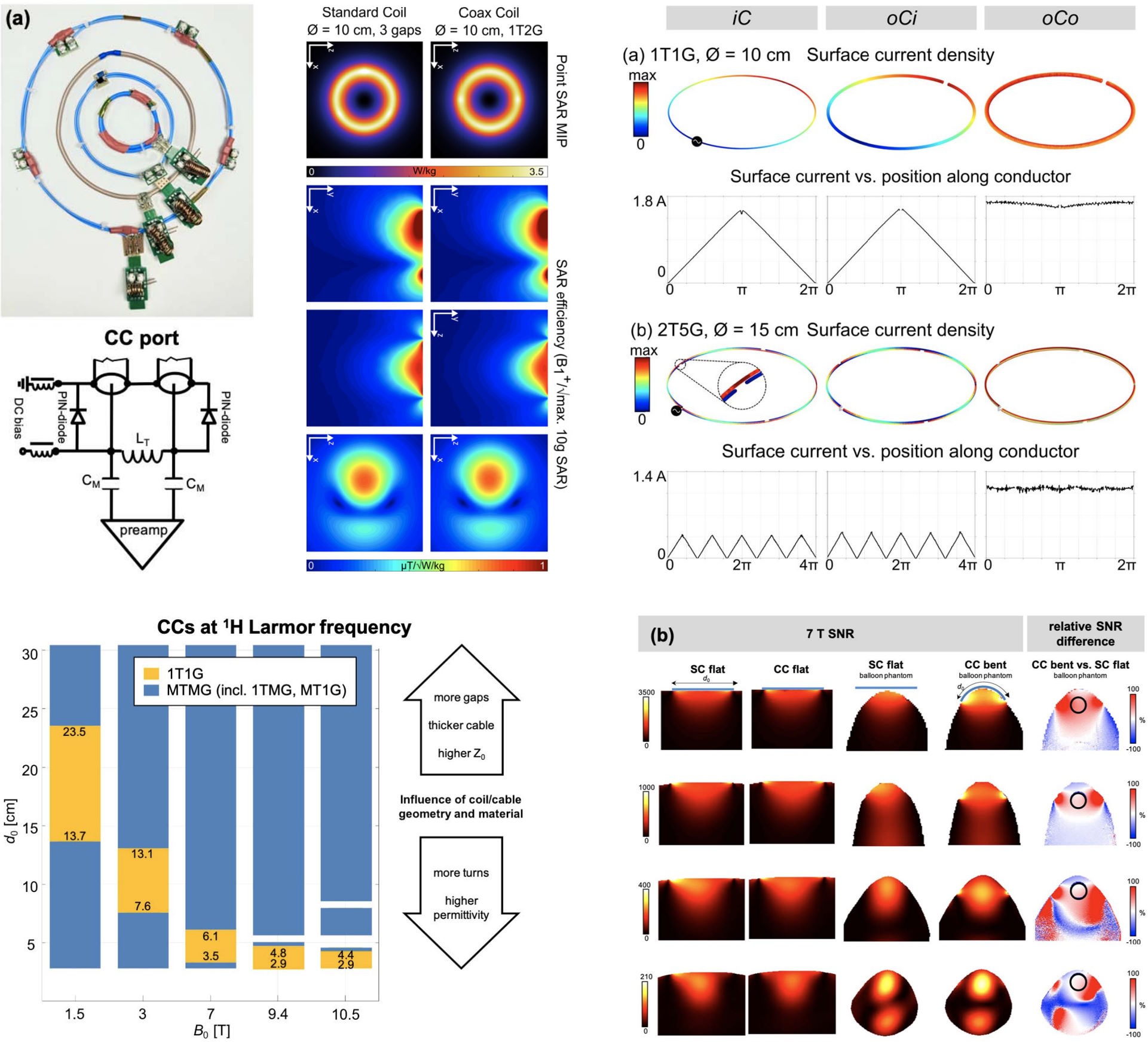
Multi-Turn Multi-Gap (MTMG) Transmission Line Resonators
Parallel plate transmission line resonators can be employed as self-resonant RF coils in MRI. Thanks to the self-resonant behavior, lumped components and soldering can be avoided, and they are perfectly suitable for fabricating flexible or superconducting coils. By introducing the multi-turn multi-gap design - in contrast to previously available single-turn multi-gap or multi-turn single-gap designs - we greatly increased the number of coil geometries to choose from for a particular target frequency and application. The proof-of-concept study was published in collaboration with our French research partners.
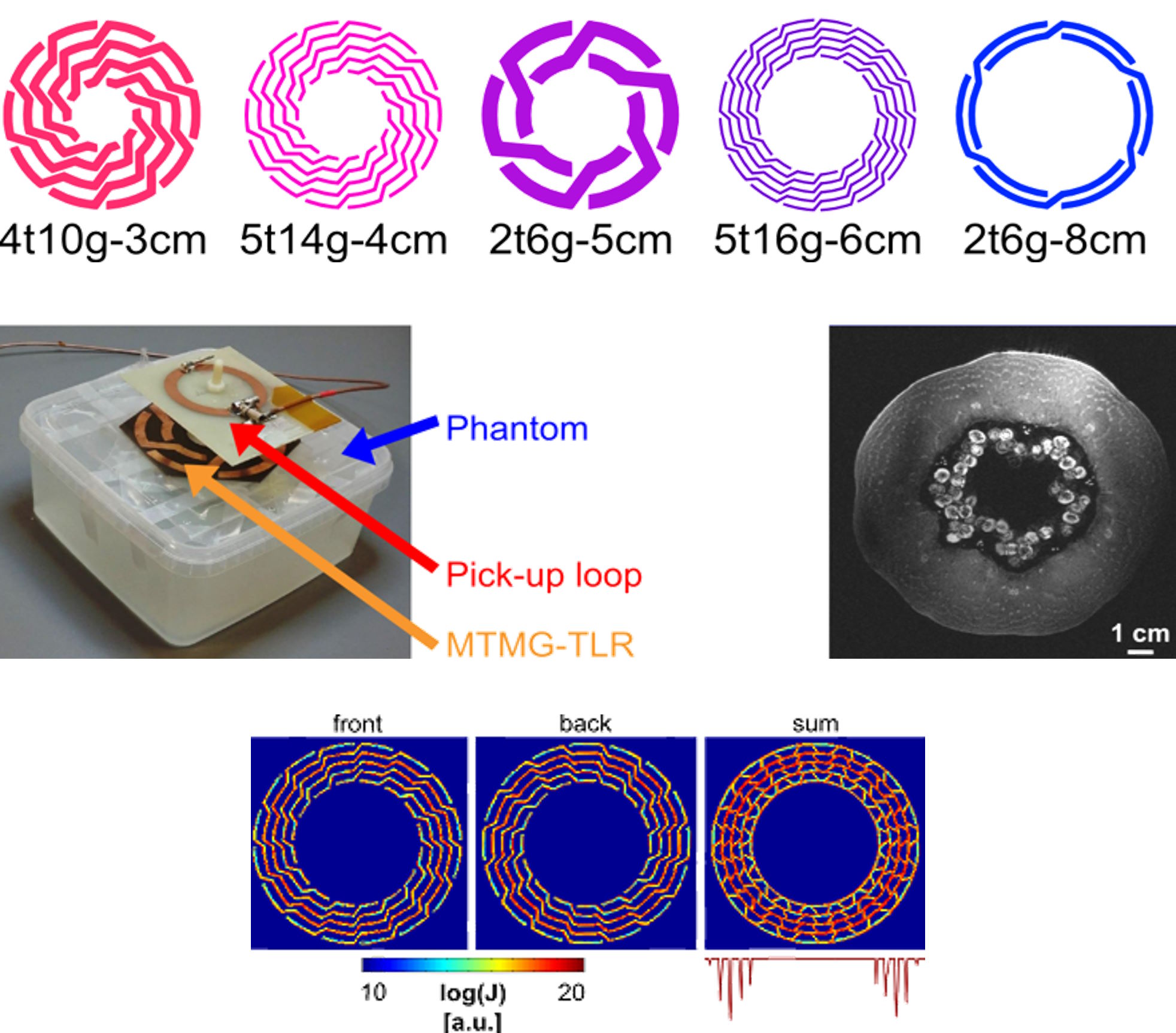
Flexible Transmission Line Resonators (TLR)
Parallel plate transmission line resonators can be employed as self-resonant RF coils in MRI. In the publication outlined below, we describe the development of the first flexible array of TLRs. The key innovation here is a novel decoupling technique based on overlapping annexes to prevent crosstalk between the individual elements of the array. To completely avoid lumped components and soldering on the TLRs, pick-up loop matching is used, which facilitates a monolithic coil design, suitable for flexible coil construction and compatible with superconducting technology.
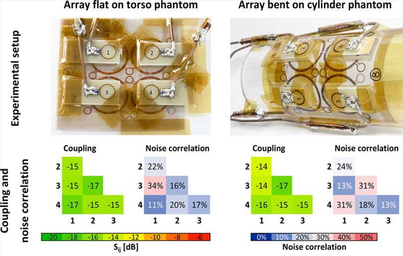
Multi-loop coils
The continuous interest in increasing the sensitivity of RF coils in MRI coined the idea of a new coil concept: the multi-loop coil. A multi-loop coil is a surface coil that is composed of multiple small loops in series. For biomedical MRI, moving from the conventional single-loop to the multi-loop design brings a sensitivity gain at high field strength (3 T and beyond). In a preliminary study performed with French collaboration partners we showed that the sensitivity gain is highest close to the coil, therefore the novel coil concept is best suited for superficial target regions, e.g. skin, cortical brain, small joints.
FlexShim: on-coil B0 shimming for flexible coils
Inhomogeneities in the static magnetic field inside the body caused by differences in magnetic susceptibility between different tissues and air result in image artifacts in MRI. To counteract these inhomogeneities, so-called shim coils are used. Typically they are built into the MR scanner bore, in some cases, additional shim coil arrays closer to the body are used. Shim coils can also be combined onto the same conductor as the RF coils (“ACDC coils”), which has been shown for rigid coil arrays. In FlexShim we investigated the integration of shim capabilities onto flexible coils and have developed a first prototype. In simulation we have shown that the homogeneity of the magnetic field within the breast could be strongly improved when equipping all RF coil elements of the BraCoil with shim functionality.

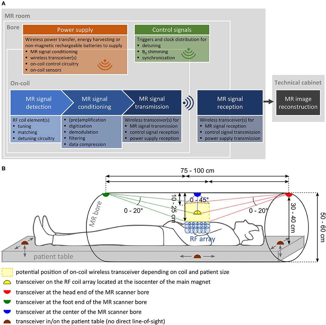
Wireless RF coils
Challenges of developing wireless radio frequency (RF) coils for MRI are summarized in one of our group’s publications. The paper delves into the requirements and strategies for three crucial components: For modern MRI setups with high-quality data demands, the need for wireless high-speed data transmission in the MR receive chain is addressed. Additionally, managing control signals and power supply for on-coil components is discussed. Entirely wireless RF coils will become a reality in the future by combining advanced scientific approaches and innovations in wireless technology, MR compatible components, and power supply solutions.
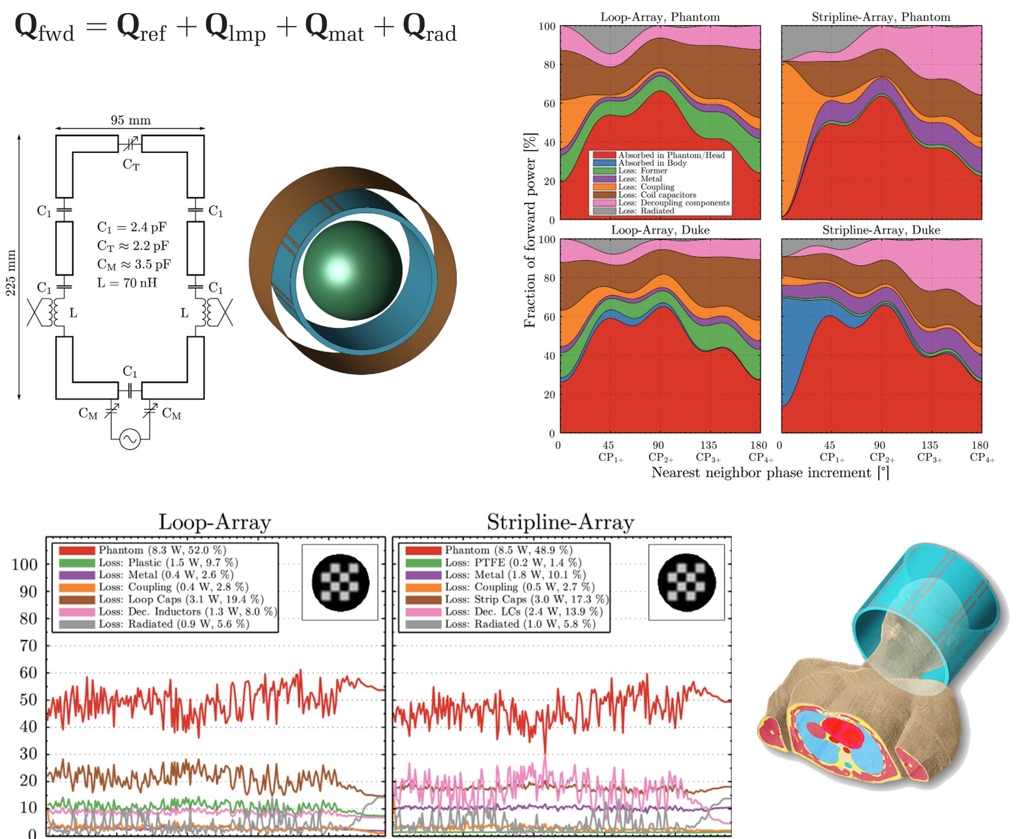
Power Balance in RF Transmit Arrays
The individual loss contributions of transmitted RF fields derived from electromagnetic simulations can be quantified using the Q-matrix formalism. This allows to investigate and optimize RF coil designs and excitation pulses, particularly for parallel transmission (pTx) at ultra-high field strength.
Research Groups
Andreas Berg | Frass-Kriegl-Group | Laistler Group | Meyerspeer Group | Schmid Group | Tik Group | Windischberger Group