Transcranial magnetic stimulation (TMS) is a non-invasive technique to induce controlled currents in cortical tissue via brief magnetic pulses.

The possibility of influencing activity in specified brain regions make TMS immensely important in the neurosciences and their clinical applications. By stimulating different targets and recording the changes in overt behavior, TMS allows creating causal maps of language- and motor function that can be used to guide brain surgery. Among other neurological and psychiatric disorders repetitive TMS has proven efficacy for the treatment of major depressive disorder.
Precision medicine approach to TMS

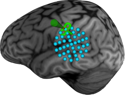
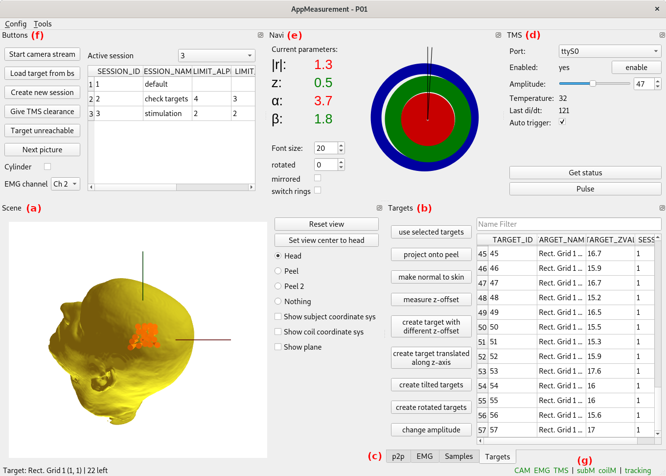
Treatment response in TMS applications can be improved by a personalised approach, in which biomarkers guide effective engagement of brain circuits. In this respect, Magnetic Resonance Imaging (MRI) can not only provide anatomical images for spatial guidance but also detect brain activation changes after or even during stimulation. We are developing new routines to personalize TMS interventions using navigation, fMRI neuroimaging and computational modelling, aiming to optimize treatment parameters including definition of stimulation targets, spatial and temporal accuracy and individualized dose.
Simultaneous TMS/fMRI

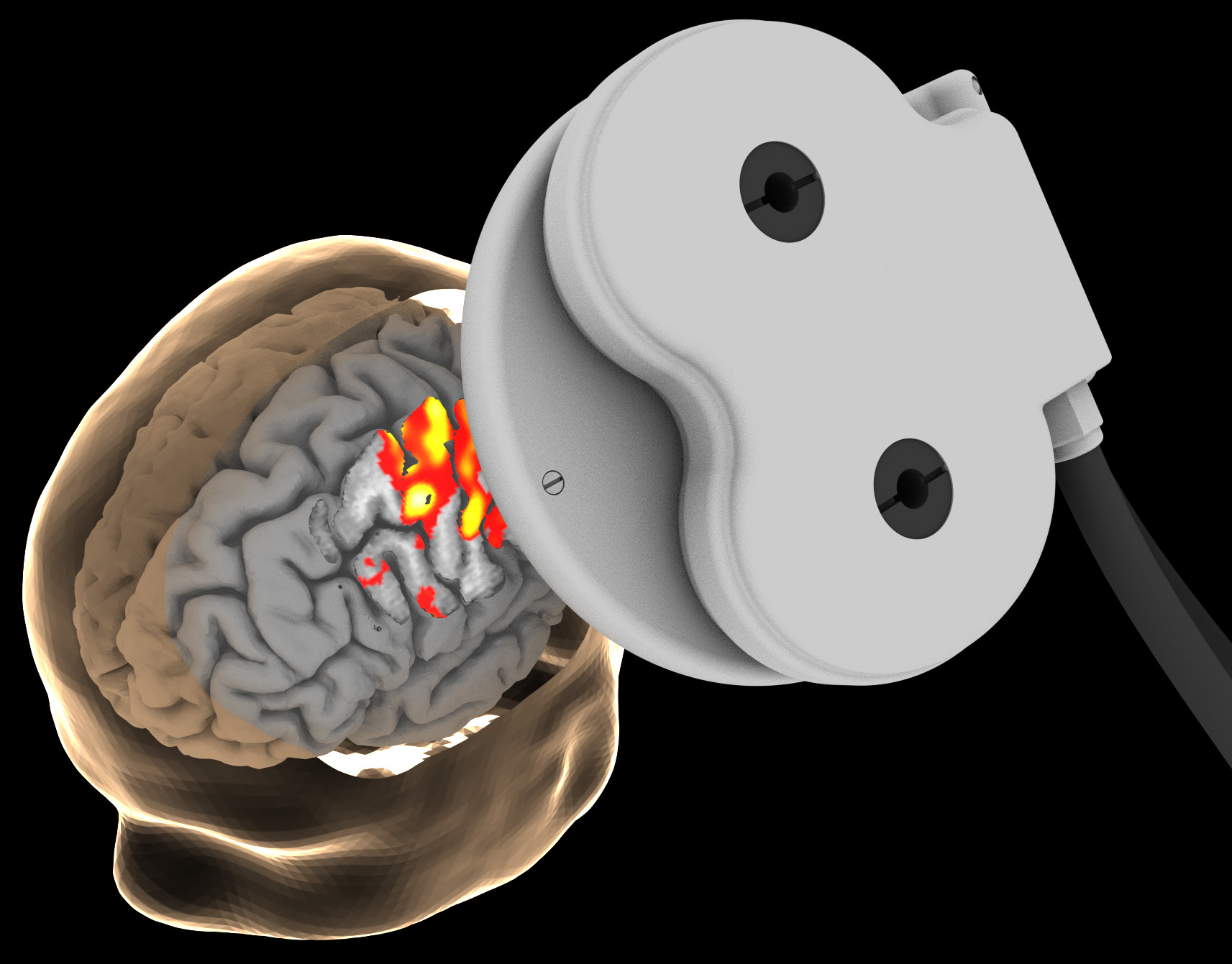
Specifically, simultaneous fMRI using a seven-channel receive magnetic resonance coil developed in-house together with the Laistler Group that is placed between the skull and the TMS coil allows to obtain fMRI data from the site of stimulation with unprecedented sensitivity. Repeated pulses of TMS, precisely controlled to avoid interference with MR allow for ensuring stimulation success monitoring.
