Damit jedes Photon zählt.

„Quantitative Bildgebung und medizinische Physik“ (QIMP) wurde 2013 gegründet. Die Gruppe ist mit einer ordentlichen Professur für „Physik der Medizinische Bildgebung – Hybride Bildgebung“ an der Med Uni Wien verbunden.
Die Gruppe besteht aus einer Vielzahl von akademischen und nicht-akademischen ExpertInnen, die durch die Absicht vereint sind, wissenschaftliche Unterstützung zur Verbesserung der Gesundheitsfürsorge für PatientInnen zu leisten. Wir engagieren uns in der angewandten Forschung, die hilft, das Konzept der personalisierten Medizin durch die Verwendung von multi-modalen Bildgebungsmethoden klinisch zu verankern.
Forschungsbereich
Gruppenmitglieder
Gruppenleiter
Thomas Beyer
T +43 (0)1 40400-39890
Mitglieder
Smaranda Bogoi
Sebastian Gutschmayer
Masar Al-Muttairi
Manuel Pires
Ivo Rausch
- Sarit Alaev
- Alexander Berger
- Matthias Blaickner
- Jacobo Cal-Gonzalez
- Zacharias Chalampalakis
- Matthew DiFranco
- S. Ehrenguber
- Amir Fatemi
- Daria Ferrara
- Marko Grahovac
- July Alejandra Gonzalez Valladares
- Kaspar Höschl
- David Iommi
- Nicole Jurjew
- Hunor Kertesz
- Corinna Kuderer
- Denis Krajnc
- Martin Lyngby Lassen
- Manuela Mayrhofer
- Sasan Moradi
- Theresa Neubauer
- Ebolyn Nwosu
- Laszlo Papp
- Nina Pötsch
- M. Raidl
- Peter Rosenbühler
- Peter M. Schaffarich (†)
- Lalith Kumar Shiyam Sundar
- Andreas Tuma
- Stefan Wampl
- Andreas Zitterl
- Ultra-low-dose PET/CT imaging (2025)
Unterstützt durch Siemens Healthcare Molecular Imaging; PI Thomas Beyer - Metabolic mapping of normative metabolism in FDG-PET (2025)
FWF (PAT 3538424), gemeinsames Projekt mit University Hospital Kanazawa, Japan; PI Thomas Beyer - HOPE-Holistic, multi-organ assessment of patient undergoing combined theranostics and systemic therapies (2024)
Unterstützt durch die ICPO Foundation; PI Thomas Beyer - MR-VIPRO - MR-Visible Plastics in Radiation Oncology (2024)
FFG Projekt; PI Ivo Rausch - MR visible polymer for medical imaging applications (2022)
Unterstützt von AWS (Austria Wirtschaftsservice) - FDG-PET Imaging for cancer-induced cachexia in lung cancer (2022)
Unterstützt von EraPerMED - Inter-organ communication assessed by FDG-PET in breast cancer patients (2022)
Unterstützt von EraPerMED - Normative database of FDG-PET images and applications in cachexia (2021)
Unterstützt von Siemens Healthineers - Improving Clinical PET imaging for non-optimal positron emitters (2018)
Unterstützt von Siemens Healthineers (SMS) - Low-count-high-quality reconstructions for PET and SPECT (2017)
Unterstützt von Fonds zur Förderung der wissenschaftlichen Forschung (FWF) - Innovative Training Network towards raising and supporting the next generation of creative and entrepreneurial cross-specialty imaging expert (2017)
EU Project 764458 “HYBRID” (H2020-MSCA-ITN-2017) - Personalized diagnosis of non-lesional epilepsy using simultaneous PET/MR (2016)
Unterstützt von Fonds zur Förderung der wissenschaftlichen Forschung (FWF) - [18F]FDG and [18F]NaF-PET Imaging of Vulnerable Plaque (2016)
Unterstützt von Siemens Healthineers (SMS) - Quantification of bone lesions in combined PET/MR and Dual-time point imaging of lesions with PET/MR (2014)
Unterstützt von Siemens Healthcare (SMS) - Multi-centre PET/CT: Quality control, FDG-guidelines and quantification (2013)
Unterstützt von the Österreichische Gesellschaft für Nuclearmedizin (OGN)
Ausgewählte Publikationen
- D Ferrara, EM Abenavoli, T Beyer, S Gruenert, M Hacker, S Hesse, L Hofmann, S Pusitz, M Rullmann, O Sabri, R Sciagrà, LKS Sundar, A Tönjes, H Wirtz, J Yu, and A Frille. Detection of cancer-associated cachexia in lung cancer patients using whole-body [18F]FDG-PET/CT imaging: A multi-centre study. J Cachexia, Sarcopenia and Muscle; 15: 2375–2386 (2024)
- LKS Sundar, et al. , Fully Automated Image-Based Multiplexing of Serial PET/CT Imaging for Facilitating Comprehensive Disease Phenotyping. J Nucl Med. 2025 Sep 18:jnumed.125.269688.
- D Ferrara, LKS Sundar, Z Chalampalakis, BK Geist, D Gompelmann, S Gutschmayer, M Hacker, H Kertesz, K Kluge, M Idzko, W Langsteger, J Yu, I Rausch and T Beyer. Low-dose and standard-dose, whole-body [18F]FDG-PET/CT imaging: Implications for healthy controls and lung cancer patients. Front Physics, Med Phys Imag. as been approved for production and accepted for publication in Frontiers in Physics, section Medical Physics and Imaging. 12 (2024)
- Sundar LKS, Muzik O, Buvat I, Bidau L and Beyer T. Potentials and caveats of AI in hybrid imaging, METHODS, 188: 4-29, 2021. PMID: 33068741
- Sundar LKS, Gutschmayer S, Maenle M, and Beyer T. Extracting value from total-body PET/CT image data - the emerging role of artificial intelligence, Cancer Imaging, 24:51, 2024
- Shiyam Sundar L. K. et. al., Whole-Body PET Imaging: A Catalyst for Whole-Person Research? J Nucl Med. 2023 Feb;64(2):197-199.
- Shiyam Sundar, L. K. et. al. Fully-automated, semantic segmentation of whole-body 18F-FDG PET/CT images based on data-centric artificial intelligence, J Nucl Med. Jun 2022,
- Rausch, I., et al. Standard MRI-based attenuation correction for PET/MRI phantoms: a novel concept using MRI-visible polymer. EJNMMI Phys 8, 18 (2021).
- Rausch, I. et. al., 2019. Performance evaluation of the Vereos PET/CT system according to NEMA NU2-2012 standard. J Nucl Med 60(4): 561-567
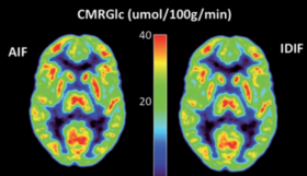
- Shiyam Sundar, L. K. et. al., 2019 The promise of fully-integrated PET/MR imaging: Non-invasive clinical quantification of cerebral glucose metabolism. J Nucl Med, 61 no. 2 276-284
- Beyer T et al., 2020. What scans we will read: Imaging instrumentation trends in clinical oncology, Cancer Imaging,
- Valladares, Alejandra,Standardized quality assurance protocols in multi-parametric imaging, PhD thesis, Medical University of Vienna, 2021.
- Kertesz, Hunor, Low-count-high-quality reconstruction methods for PET and SPECT imaging, PhD thesis, Medical University of Vienna, 2022.
- Papp, Laszlo, Computer-aided image-derived tumour characterization utilizing multi-layer machine learning, PhD thesis, Medical University of Vienna, 2021.
- Shiyam Sundar, Lalith Kumar, Personalized neuroimaging for improved assessment of non-lesional epilepsy based on fully-integrated PET/MRI imaging, PhD thesis, Medical University of Vienna, 2019.
- Rausch, Ivo, Advanced quantification for fully-integrated PET/MRI : implementing MRI-based attenuation correction in clinical routine, PhD thesis, Medical University of Vienna, 2017
- Lassen, Martin Lyngby, PET Quantification in PET/CT and PET/MR, PhD thesis, Medical University of Vienna, 2017


